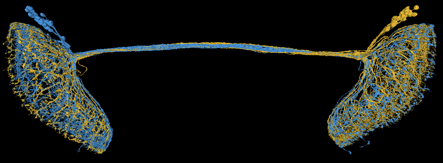
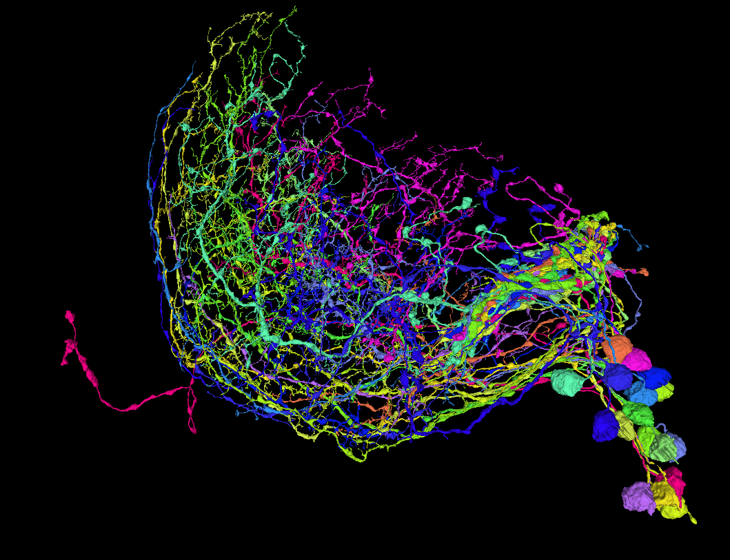
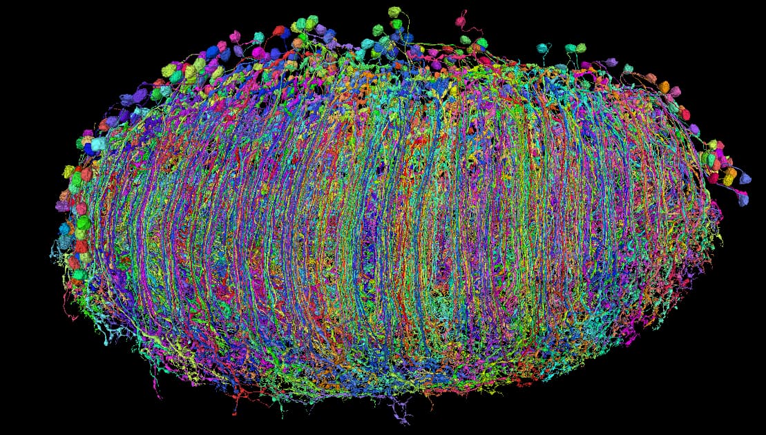
A bed of kelp?
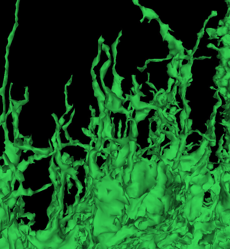
Or something eldritch rising from the deep?
(featured from this chunk of glia and friends)
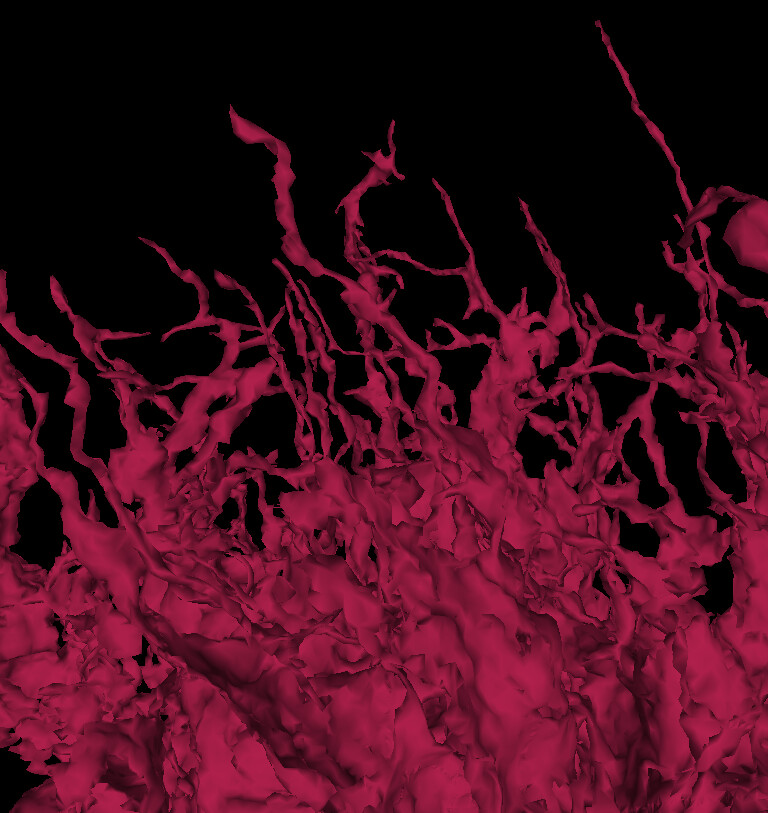
A bed of kelp?
Or something eldritch rising from the deep?
(featured from this chunk of glia and friends)
Nice fan/snow shovel made by LC14b cells in the left optic lobe:
https://ngl.flywire.ai/?json_url=https://globalv1.flywire-daf.com/nglstate/4989239164928000
And LC14 cells themselves:
https://ngl.flywire.ai/?json_url=https://globalv1.flywire-daf.com/nglstate/5652828120940544
Gorgeous!
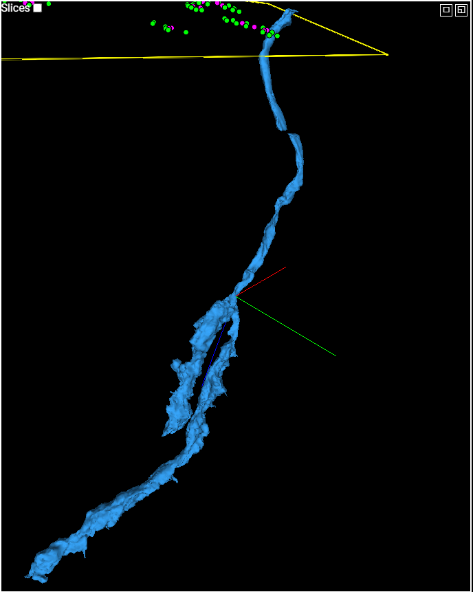
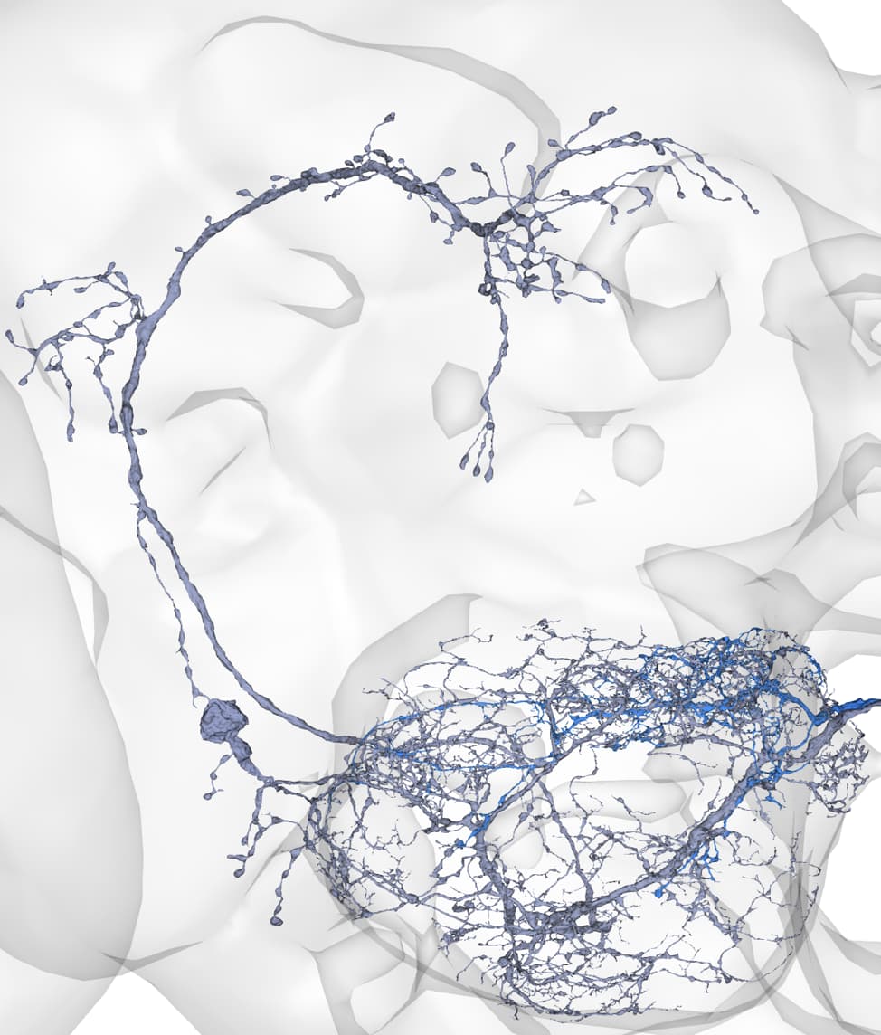
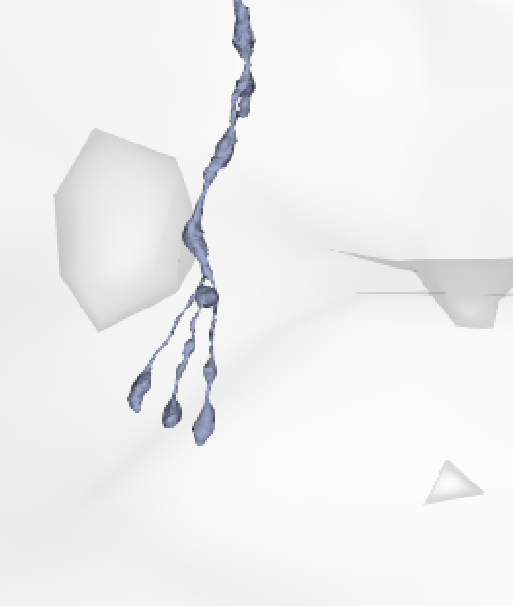
an interesting (almost) double barrelled R1-6
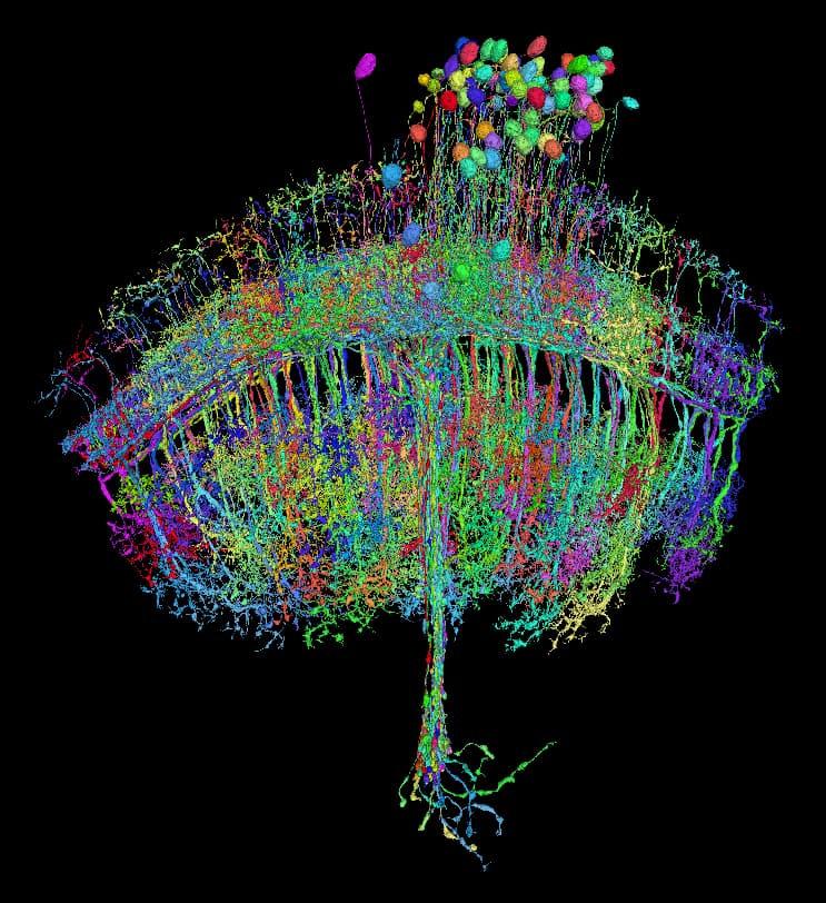
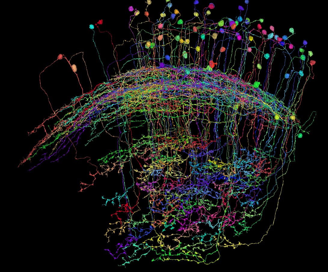
Interesting collection of the TmY14 cells. It seems, that these are almost all of the cells in this lobe, yet the somas are only in this relatively narrow band of the medulla cortex.
The TmY14 cells are also unique, that they are the only Tm/TmY cells, that project into the central brain.
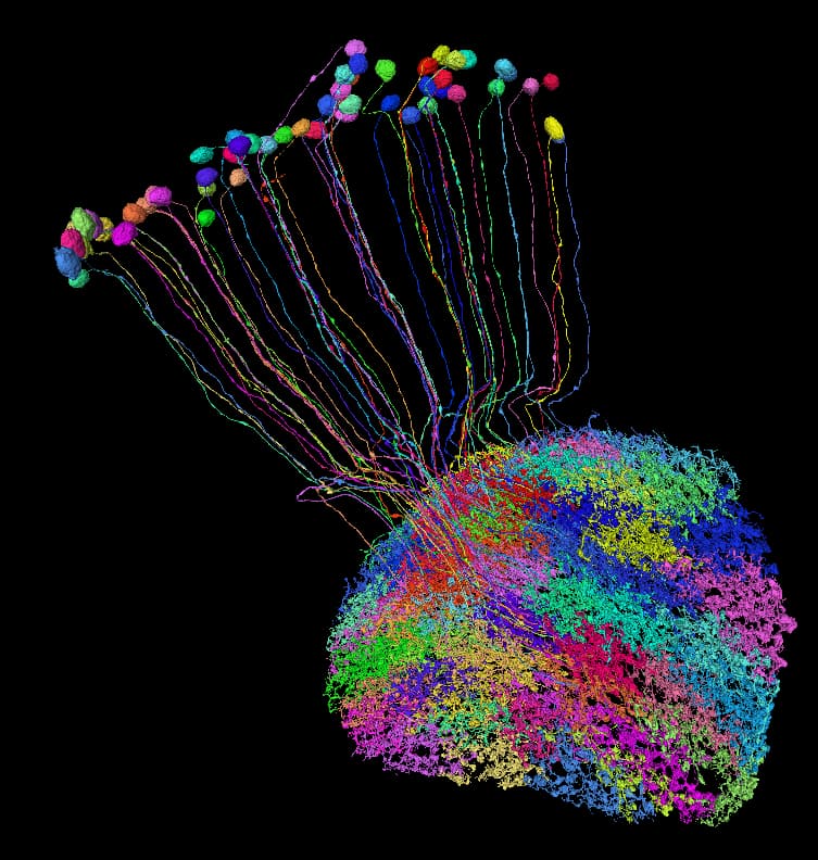
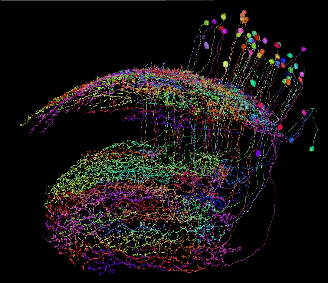
Interesting bunch of cells:
It’s probably some unknown type of LPi or LPt.
The interesting thing is, that they form a glomerulus, which would be a first in the optic lobe (lobula plate, to be exact) if I’m not mistaken.
Wow that’s a pretty curious arrangement!
oooo do you mind if I share this with an optic lobe researcher? If they are new I nominate them to be named the Kruk cells!
gorgeous!
Yes, sure!
That would be so cool! ![]()
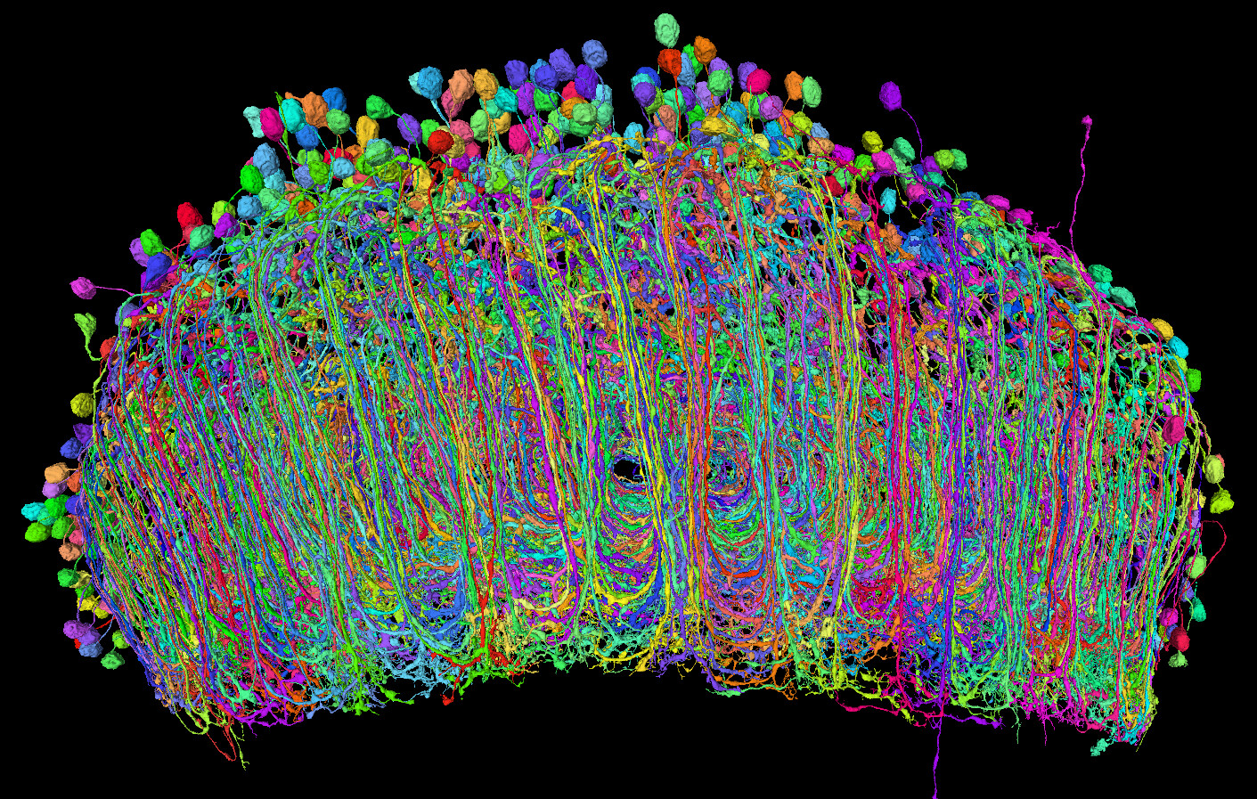
I was able to find quite a few new T4 and T5 cells in the left optic lobe (new, as in “unlabeled”, not as in “a new type”). When I’ve looked at them (T5b in the example below) from the Medulla’s perspective, it’s so cool to clearly see the channels between them, despite that there are over 700 cells in this picture:
Edit:
And these channels are for all the T4 cells (T4b in the picture): (it’s better visible in 3D, when you rotate the cells)
Nicely visible layers of Y1 (orange), Y11 (green) and Y12 (blue) in Lobula Plate. Also visible zones, where each groups’ cells’ somas are:
Wow those really are amazing cells
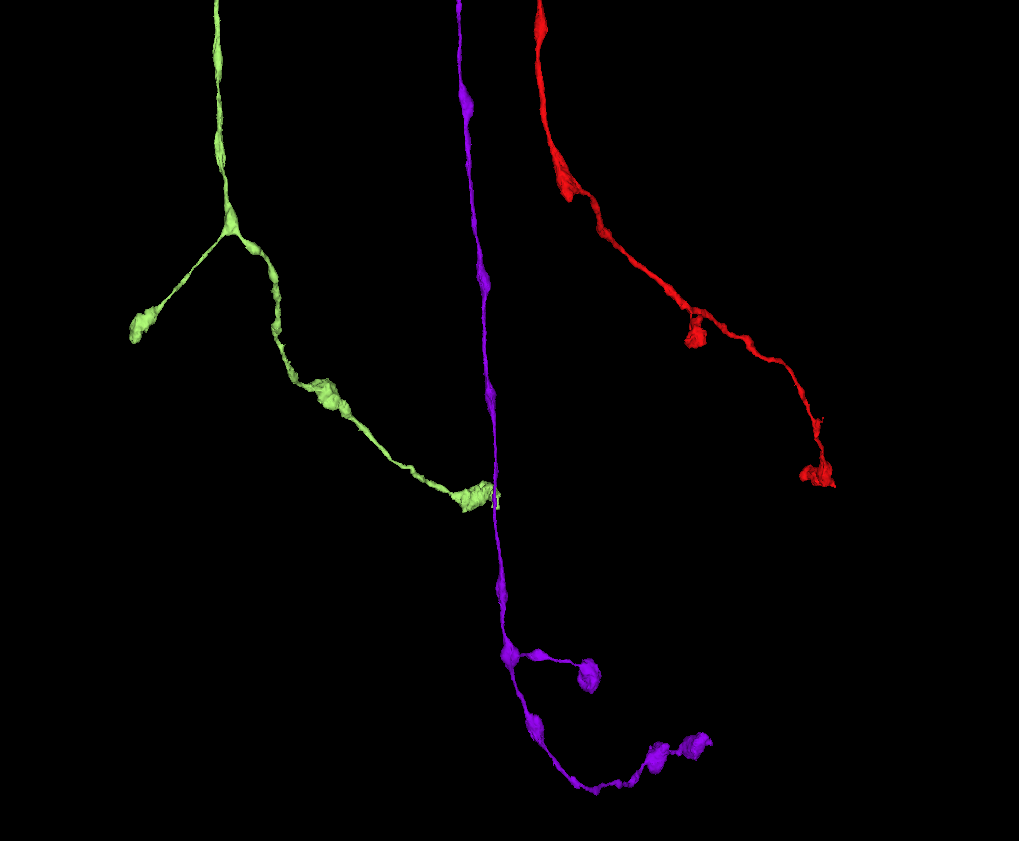
A word of caution, when working with TmY cells:
All three fragments on the picture below, are branches of the same type - TmY3. All TmY types are characterized by they extension to the lobula plate (as opposed to the Tm types). The light green on the picture indeed has such extension. However, the violet one has the extension going in reverse order - back to the lobula and the red one doesn’t have such branch at all, just a small hump.
One could say, these are some Tm cells. At this moment, I’ve checked probably tens of thousands of Tm and TmY cels and the rest of the pictured cells is quite characteristic for this one type and I didn’t see anything similar, that could be another type, so I’m 99,9% sure, these are indeed TmY3.
interesting are the violet and red ones anomalies or are there more cells with this characteristics in the dataset.
Is there any chance there could be a merger somewhere upstream so the top part is TmY3 and the bottom another type of cell?
There are a couple more like the red and violet ones and over 300 of the “normal” type.
I’ve checked them both for mergers and missing parts, but didn’t find anything.
I don’t think, there’s a merger between the top and the bottom part, because the bottom parts looks perfectly ok, other than that small branch being different. There aren’t any (afaik) other types with extactly this version of the lobula branch.
I’m also seeing similar variabilities in other TmY types. So, I guess, biology isn’t an exact science and it likes to change some stuff, if needed.
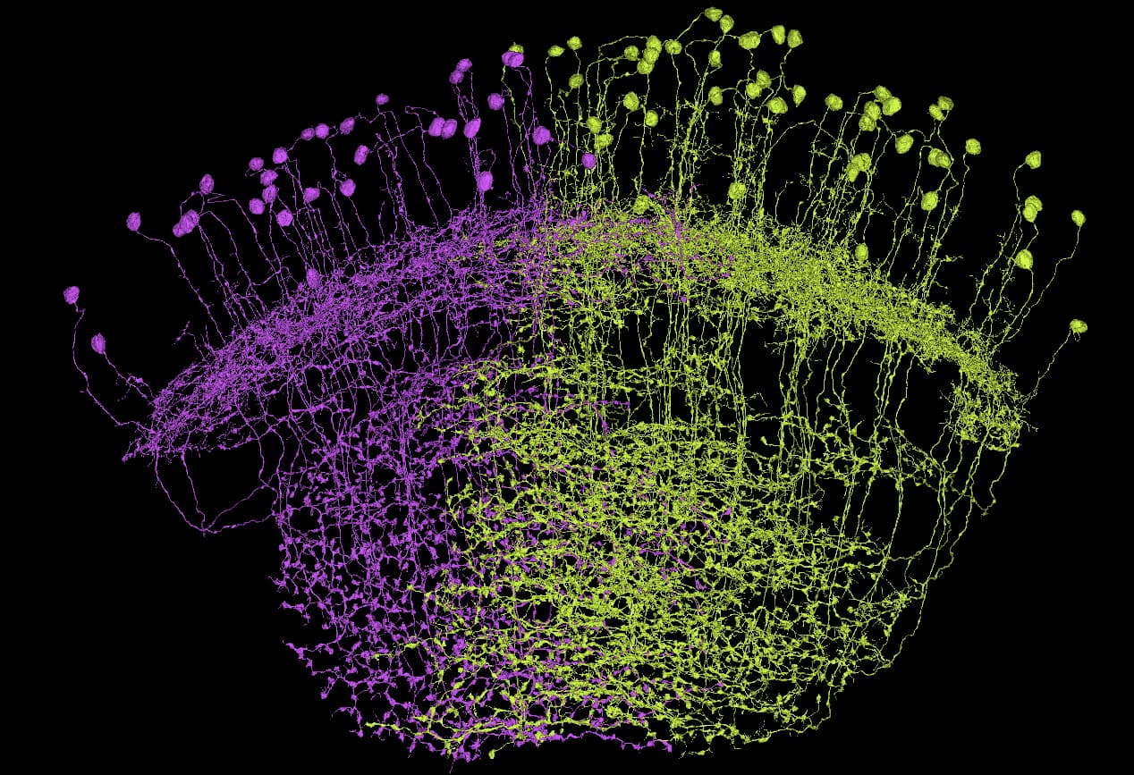
Quite unusual pair of Tm types:
https://ngl.flywire.ai/?json_url=https://globalv1.flywire-daf.com/nglstate/5659446548103168
Both types are probably unknown as of now.
Two interesting things about them are:
I’ve added them to the google sheets as “unknown 2a” (violet) and “unknown 2b” (lime) in the “Tm” section.
Edit:
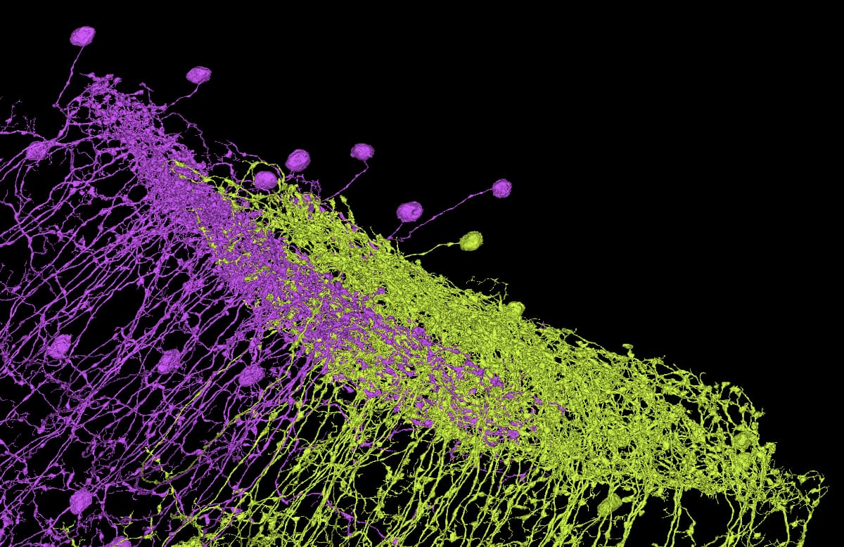
Also a bit atypical new (?) Tm type:
Atypical, because all the cell bodies are on one side of the lobe and all the arborizations grow in the other direction.
Marked as “unknown 3” in the sheet.
Edit 2:
Another similar group, but this time, the layer in the medulla splits in two:
“unknown 4” in the sheet.