Something to keep in mind when proofreading in or near the right optic lobe -
There is an area in this lobe where the lamina has partially detached from the medulla. Proofreading can be tricky in this area and sometimes there may not be enough visual information to complete a cell you’re working on.
If you have a cell that appears to end without completion please check the EM image to see if you may be stuck in this area.
There is currently not a great fix for these cells, though some of them can be remedied by seeing how they interface with nearby completed cells. But being aware of your cell’s location can help you rule out other issues, and determine next steps.
Please don’t confuse these with R cells, which also leave the dataset without completion, but exit out of the lamina rather than the medulla.
Optic lobe separation:
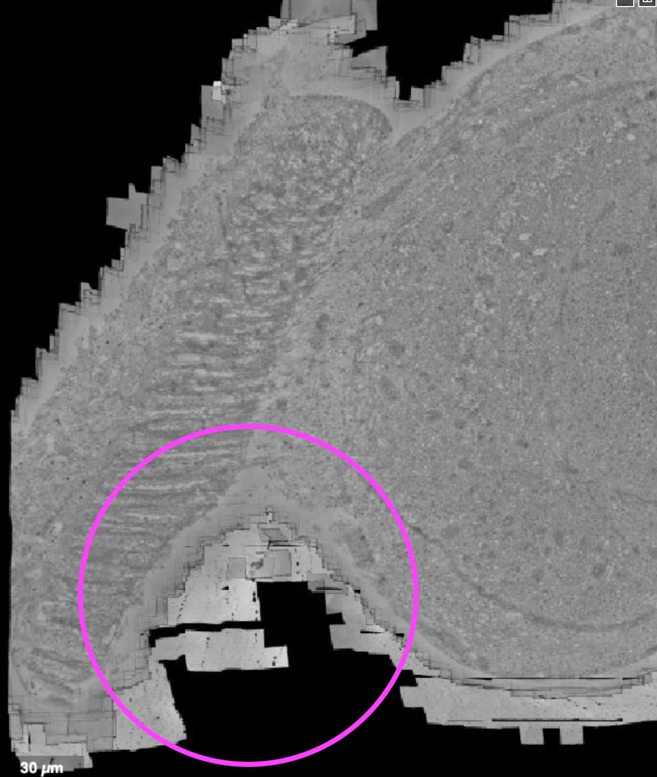
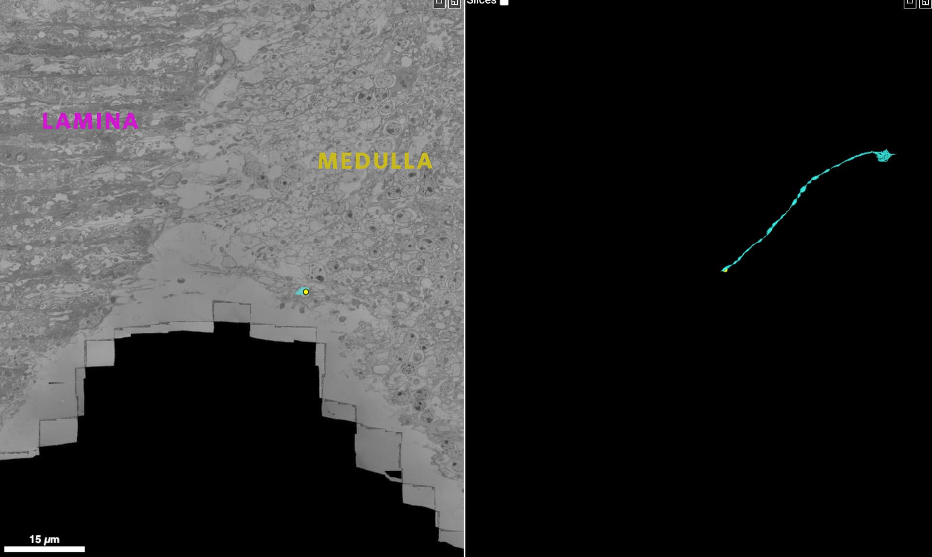
An incomplete cell due to this separation:
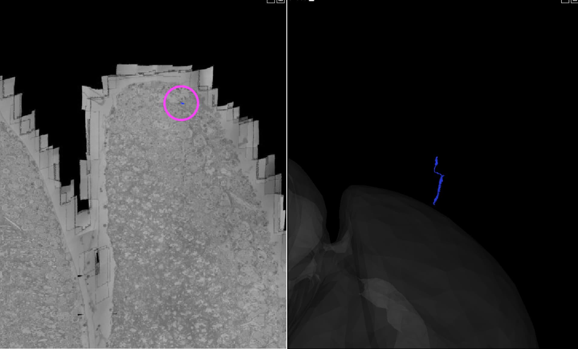
Retinula cell (R) - example of where this cell type terminates:
**Note - The right optic lobe appears on the left side and the left optic lobe on the right side when you are viewing the EM images.