Thanks! I added this to the cell deck
2 Likes
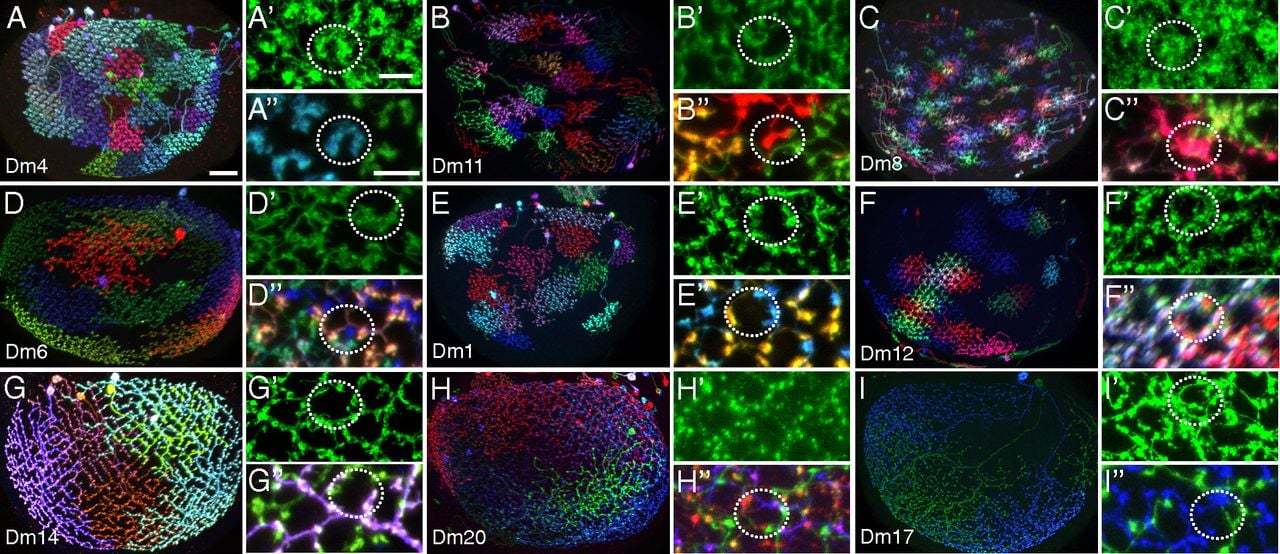
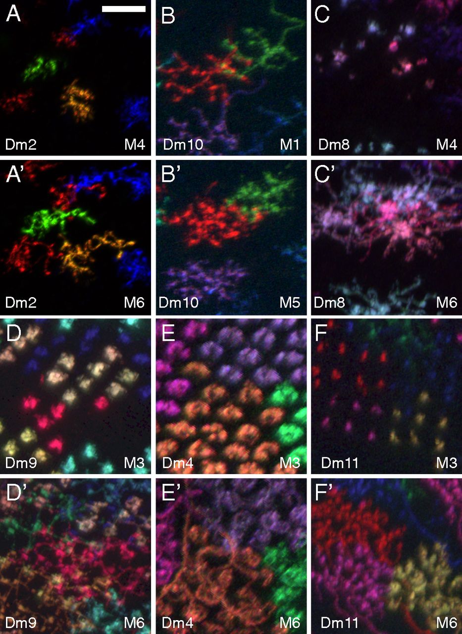
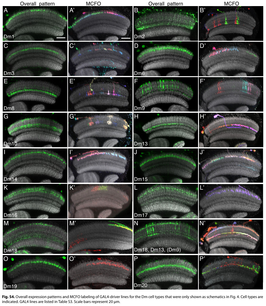
Some more images from the same source as in the first post of this thread, and its appendix:
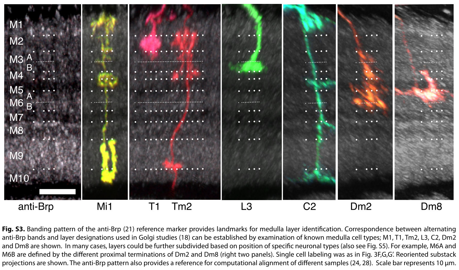
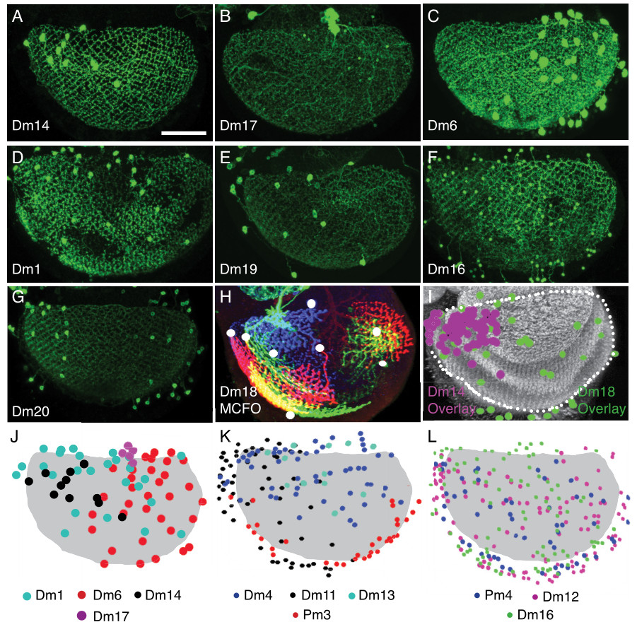
(the dots mark positions of the somas)
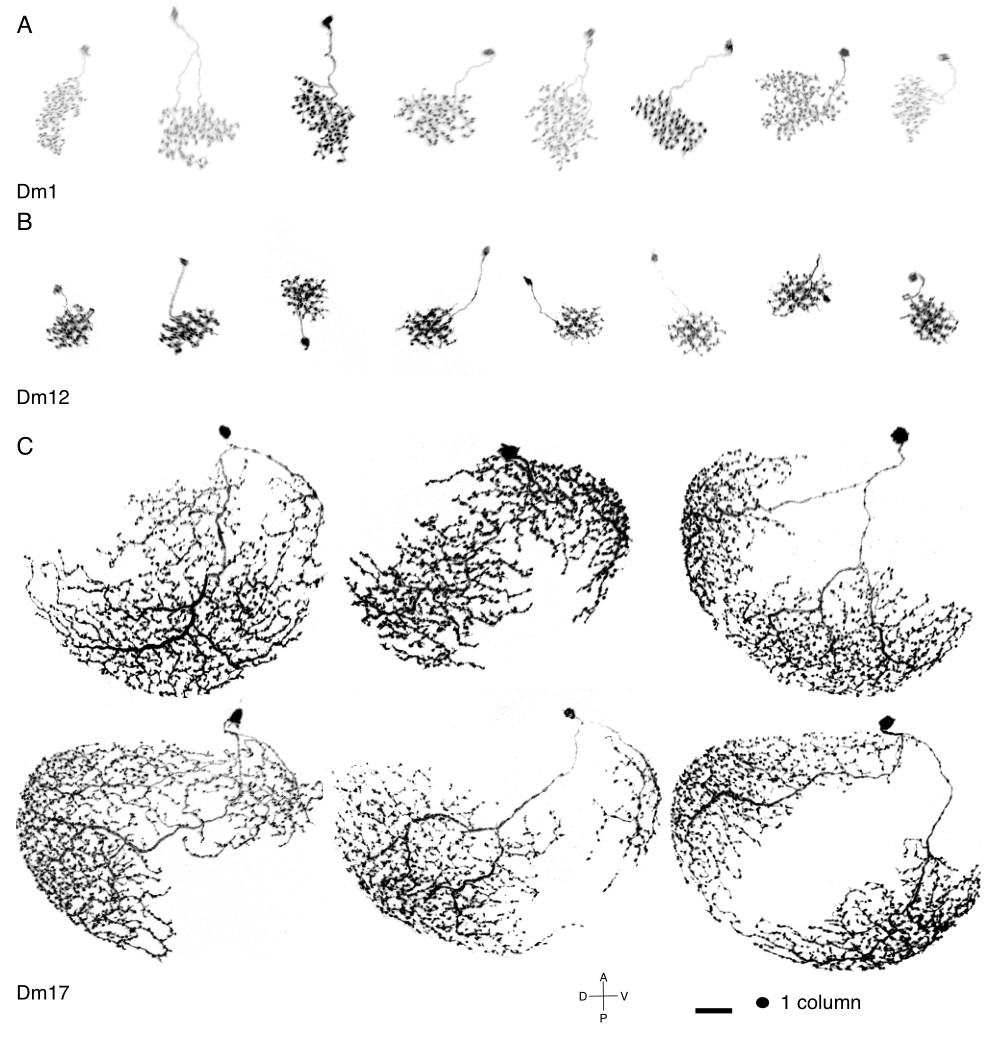
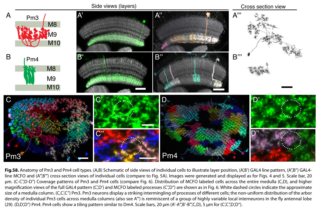
Keywords: Mi1, T1, Tm2, L3, C2, Dm1, Dm2, Dm3, Dm4, Dm6, Dm8, Dm9, Dm10, Dm11, Dm12, Dm13, Dm14, Dm15, Dm16, Dm17, Dm18, Dm19, Dm20, Pm3, Pm4
1 Like
I had trouble recognizing, which Dm is which, so I made this collection of the basic 20 types:
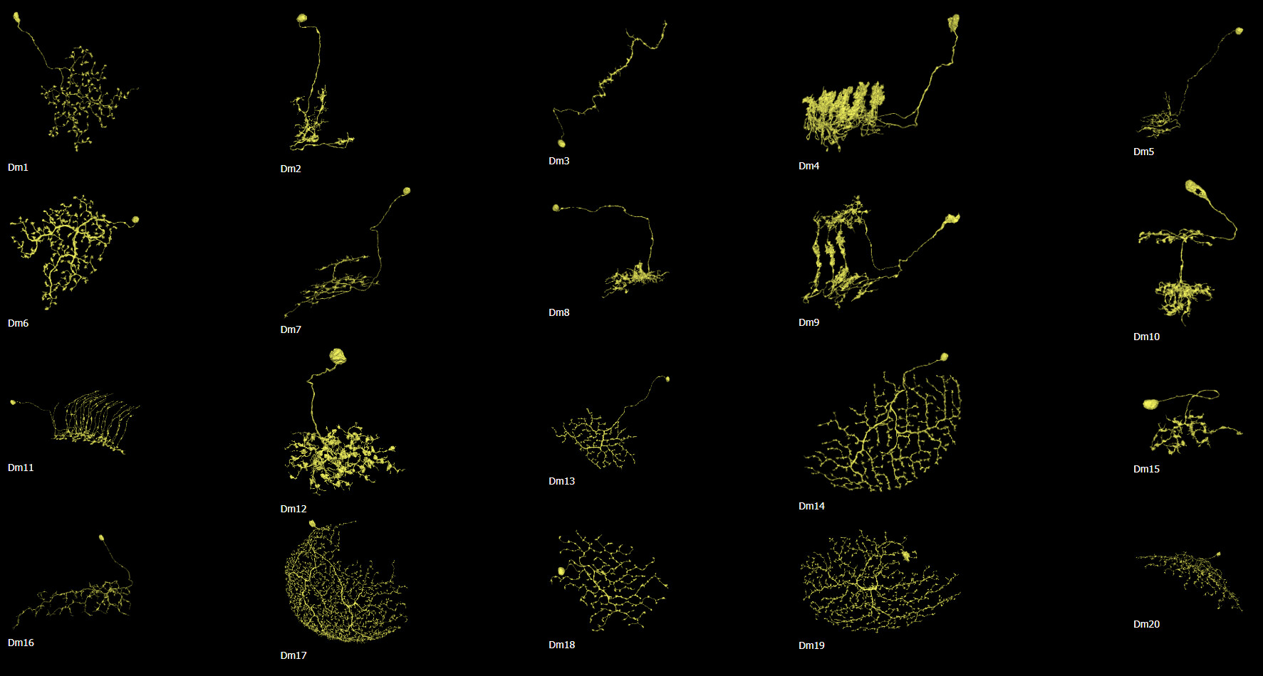
Cells are NOT up to scale to each other, because the differences would be to big and some smaller cells wouldn’t be recognizable.
5 Likes
Great visual, I added to the cell guide
1 Like
I’ve collected all the Mis from the Fischbach’s paper, scaled them, rotated, removed the original layers and put side by side in their respective layers.
Also added Mi13, Mi14 and Mi15 from this paper. These have inverted colors and set thresholds to make them black&white.
Here’s the result:

5 Likes