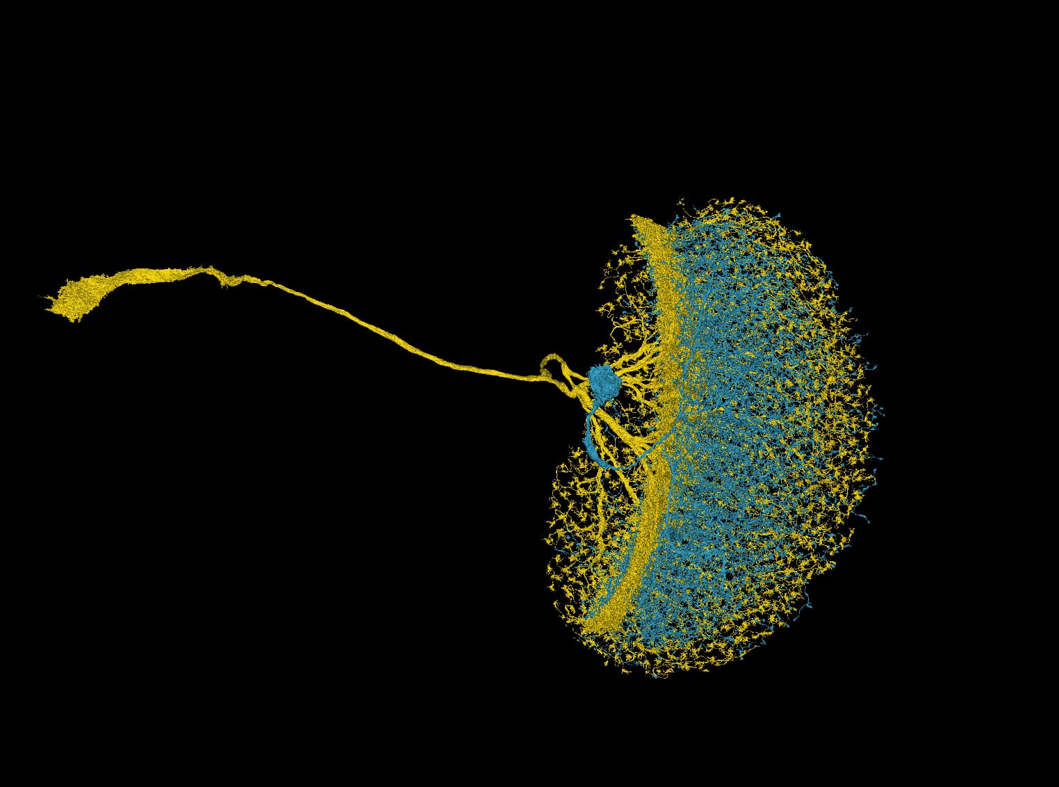
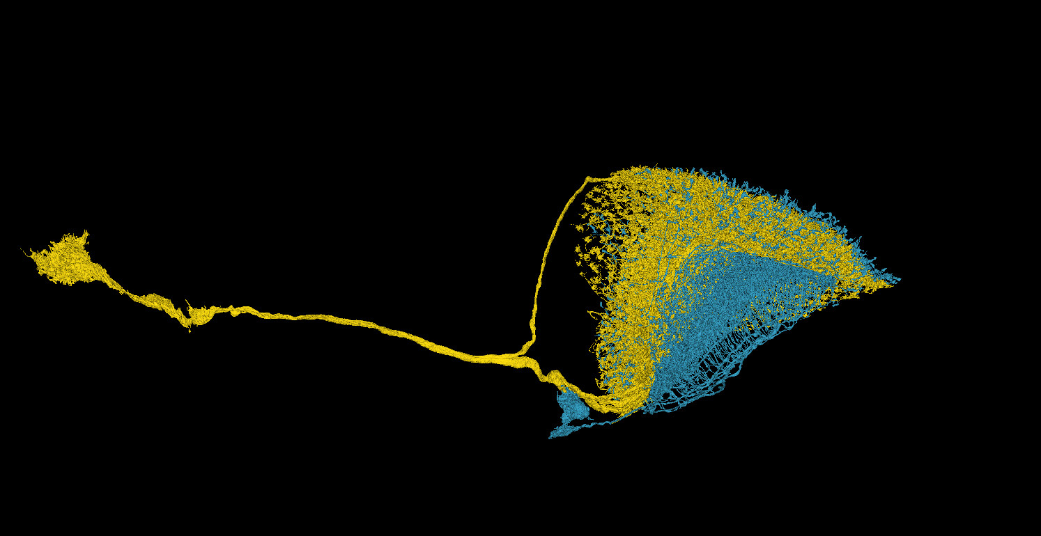
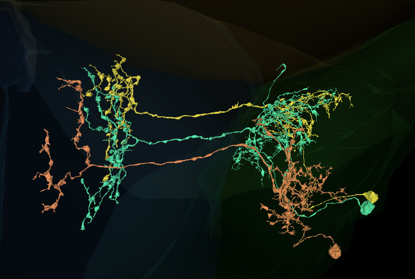
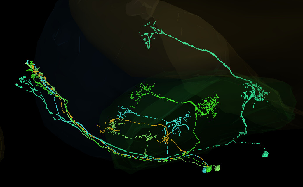
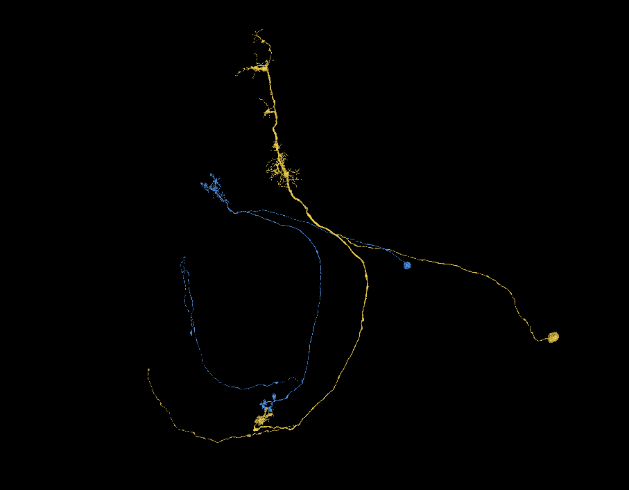
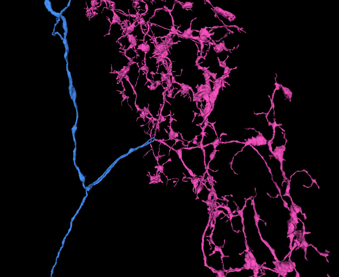
Two cells tangled and pulling to their own ways.
https://ngl.flywire.ai/?local_id=25c3b2a287fa600aaacb3e527f85e6f3
Two cells tangled and pulling to their own ways.
https://ngl.flywire.ai/?local_id=25c3b2a287fa600aaacb3e527f85e6f3
Copying a message from Kenneth Colodner:
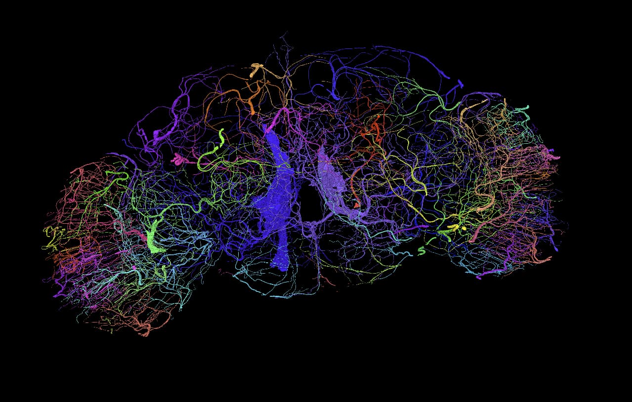
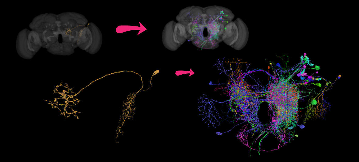
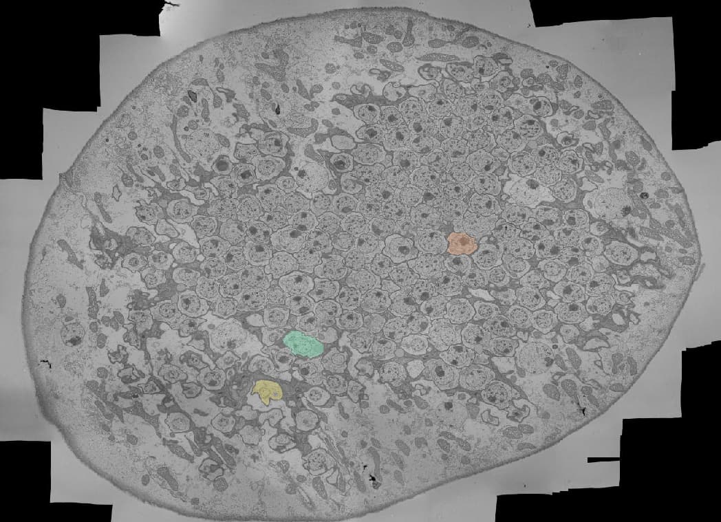
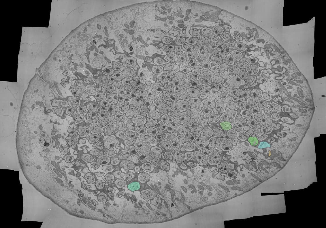
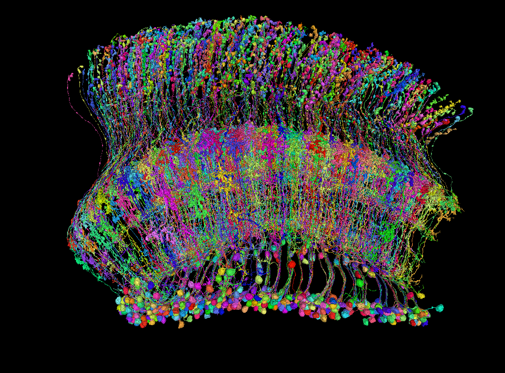
Hello FlyWire community – I’m happy to share our recent achievement. My team of undergraduates at Mount Holyoke College, led by @Mai Bui, @Sky Cho, and Marina Lin, and in collaboration with @Davi Bock, @Philipp Schlegel, and @Andrew Dacks, have proofread the entire tracheal system of FAFB in FlyWire. This is the gas exchange system for the fly, and is the identity of those tubular structures you may have encountered in your proofreading. We are at work on describing the neuroanatomical properties of this system (and will present at SfN), but I thought you would enjoy seeing what else lurks in this brain! Cheers!
<<headsup it takes a while to load!>>
So, cool! I’ve always wanted to do it, but never found a time.
This is great - I’ve loved these structures and the full map image is just as cool!
Decide to do a few Dm4’s as a break from the hallowxmas grind, and return to see this monstrosity that i never finished before:
https://ngl.flywire.ai/?json_url=https://globalv1.flywire-daf.com/nglstate/5081663281299456
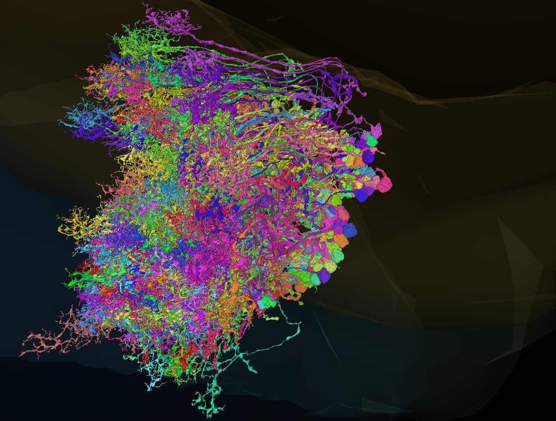
Slowly but surely, slice by slice, the outer lobula plate neurons are being sorted. This EM slice is right at the edge of the data set outside the lobula plate, and it just gets more dense the closer in it gets. I know some of you have a lot more time to work on Flywire than I do, but I’m still proud of how far I’ve come in a couple of days.
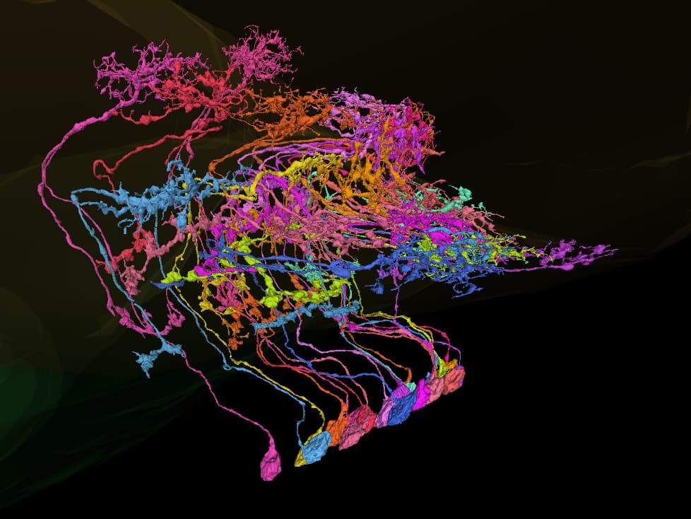
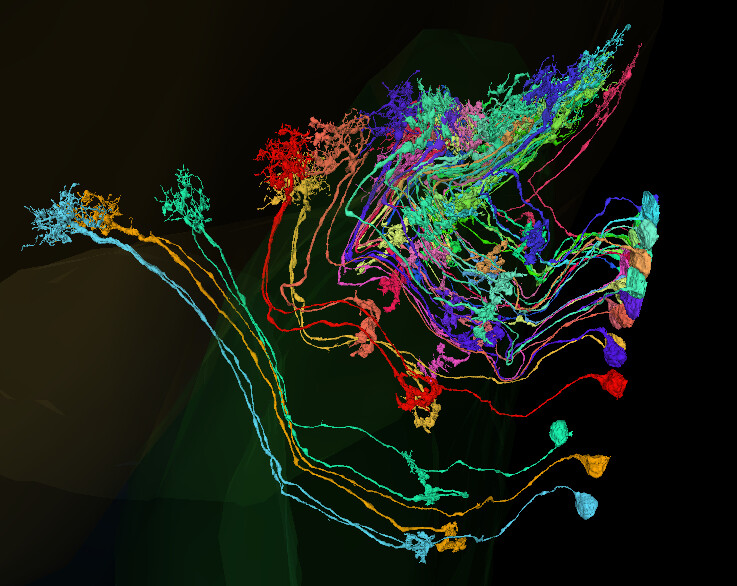
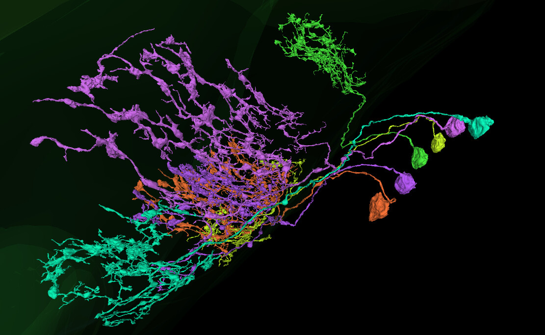
For something more colorful, have a cluster of the T4 cells I’ve found so far in this grouping:
nice farm! T2s and 5s were/are my favourites, my farm started with me wanting to find them all, lol, now its at 5.2k cells lol.
If you have a list of 5,000 cells (or even a couple hundred) that are all the same type/known type we can batch label them!
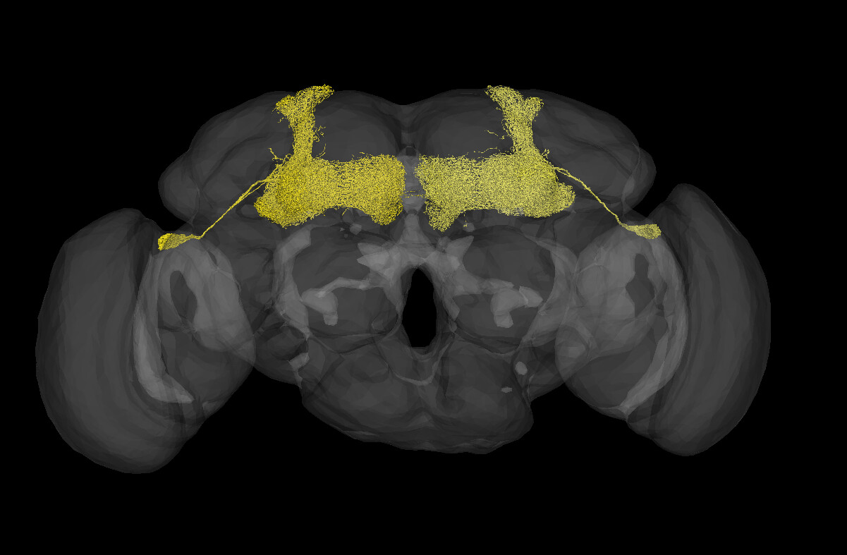
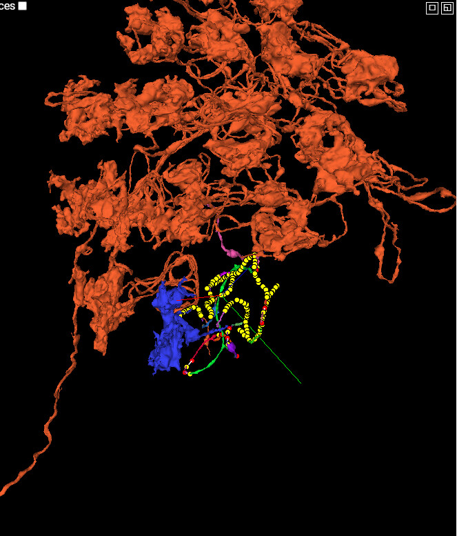
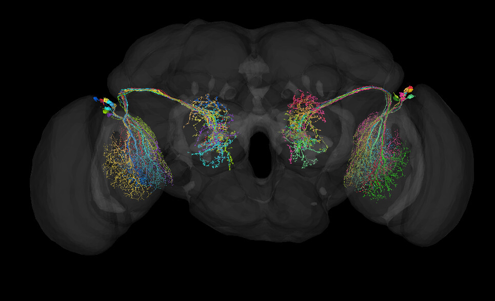
Was looking at downstream connectivity of DM8s and it went really crazy downstream from this cell - visual projection neuron labeled aSP29a. That point that the signal goes BOOM!
asp29a has such nice symmetry!
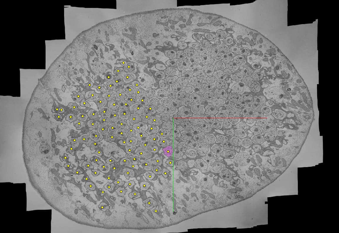
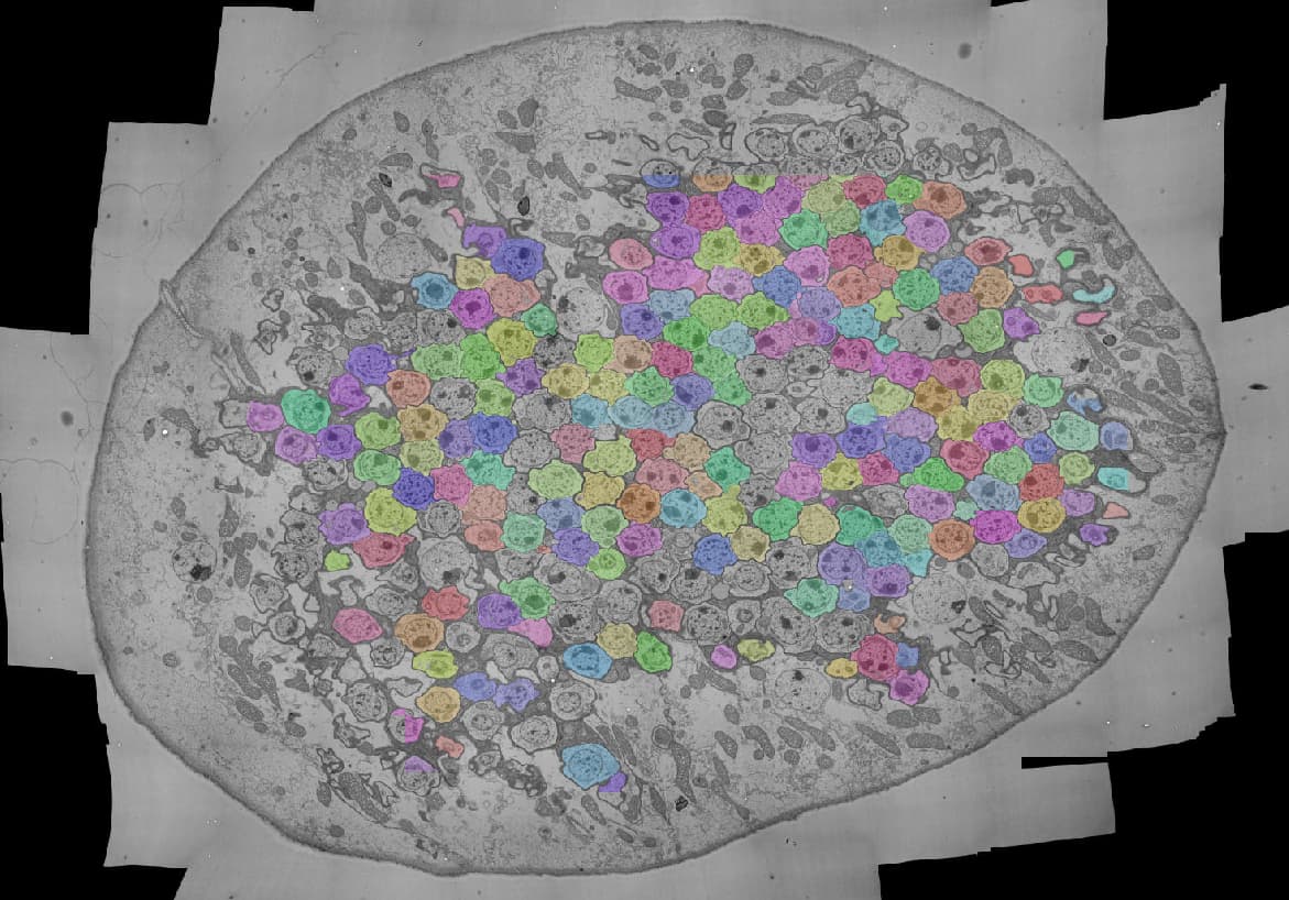
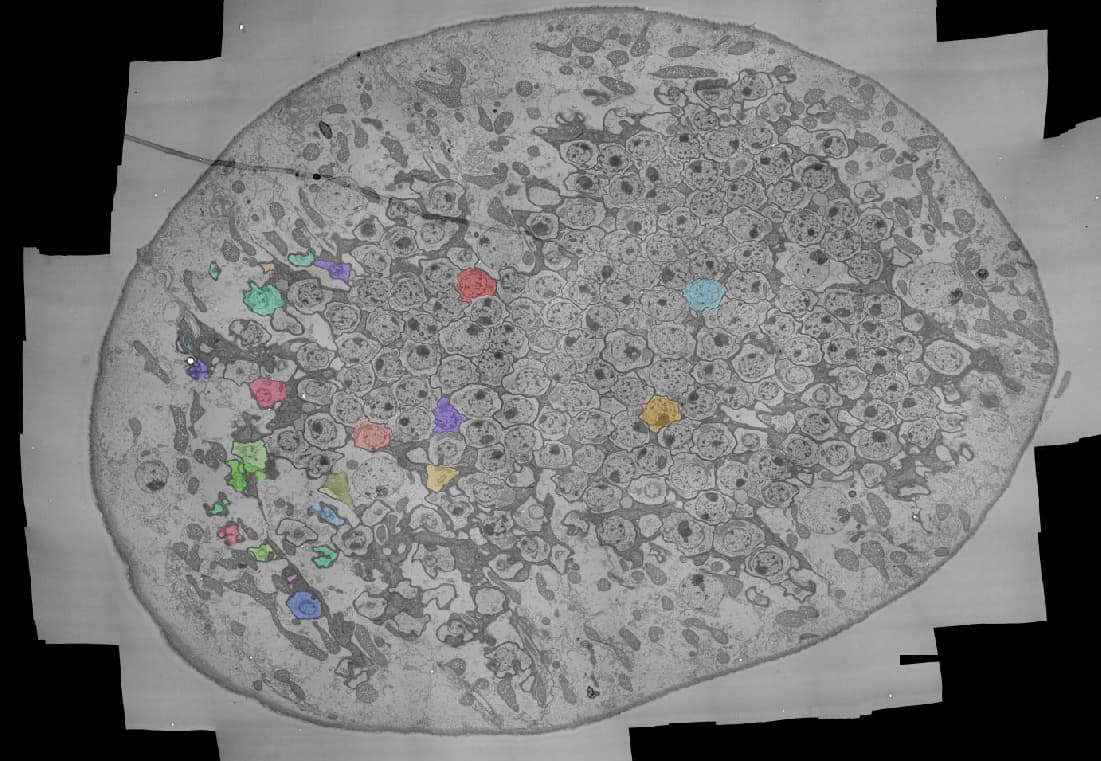
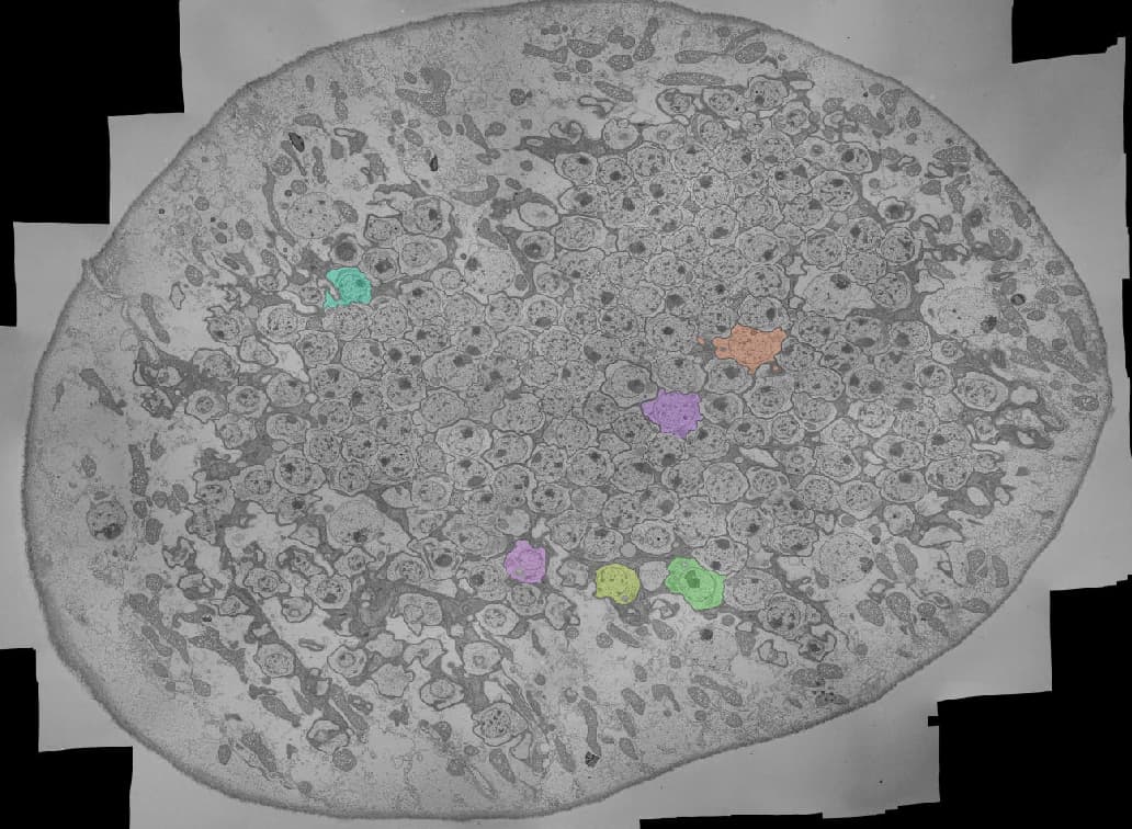
I’ve finished going through identifying and proofreading the outermost slice of the lobula plate (the lobula plate cell body rind), and thought I’d share findings and images!
As you can see here, most of the somas in this outermost range are T5 types. There are approximately 214 identified T5 cells on this layer (approximate because a few I have identified are a little further in). Link: FlyWire
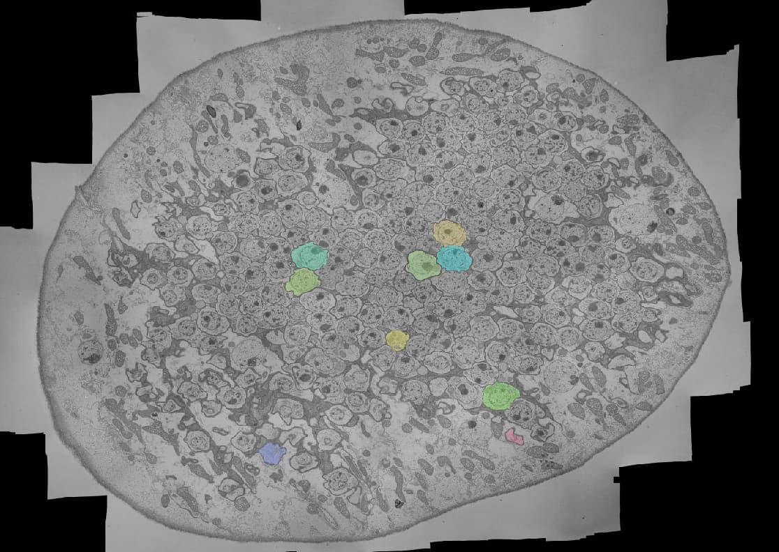
Only about 27 cells are T4 type. Link: FlyWire
About 10 are Y type. Link: FlyWire
Three are Tlp type: FlyWire
6 are Lpi type: FlyWire
And 5 are LLPC type: FlyWire
Finally, I have 8 neurons that I will post in “What Cell Is It?” that I could not classify or reasonably complete. Assistance on these would be appreciated!
That makes a total of about 271 cell types on this outer EM slice. What’s next? Well, I shuffle on up a few EM slices and start identifying the next layer! There will be a lot more somas as I head further in, but it’s interesting to see how the lobula plate exterior is forming.
This is incredible, AzureJay! It must be so satisfying to have completed that whole layer ![]()
It is so much! Even if it’s only the beginning, it’s so good to see a full slide processed.
Wow this is so cool!
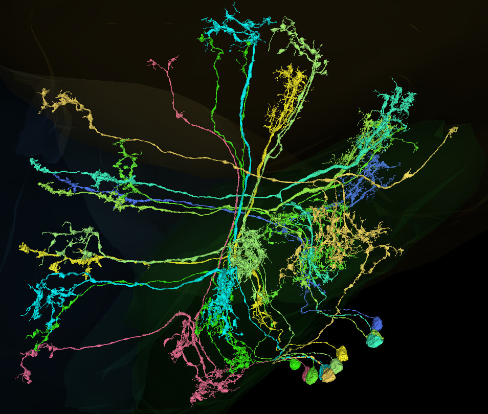
All the C cells, I’ve completed:
https://ngl.flywire.ai/?json_url=https://globalv1.flywire-daf.com/nglstate/4777855682609152
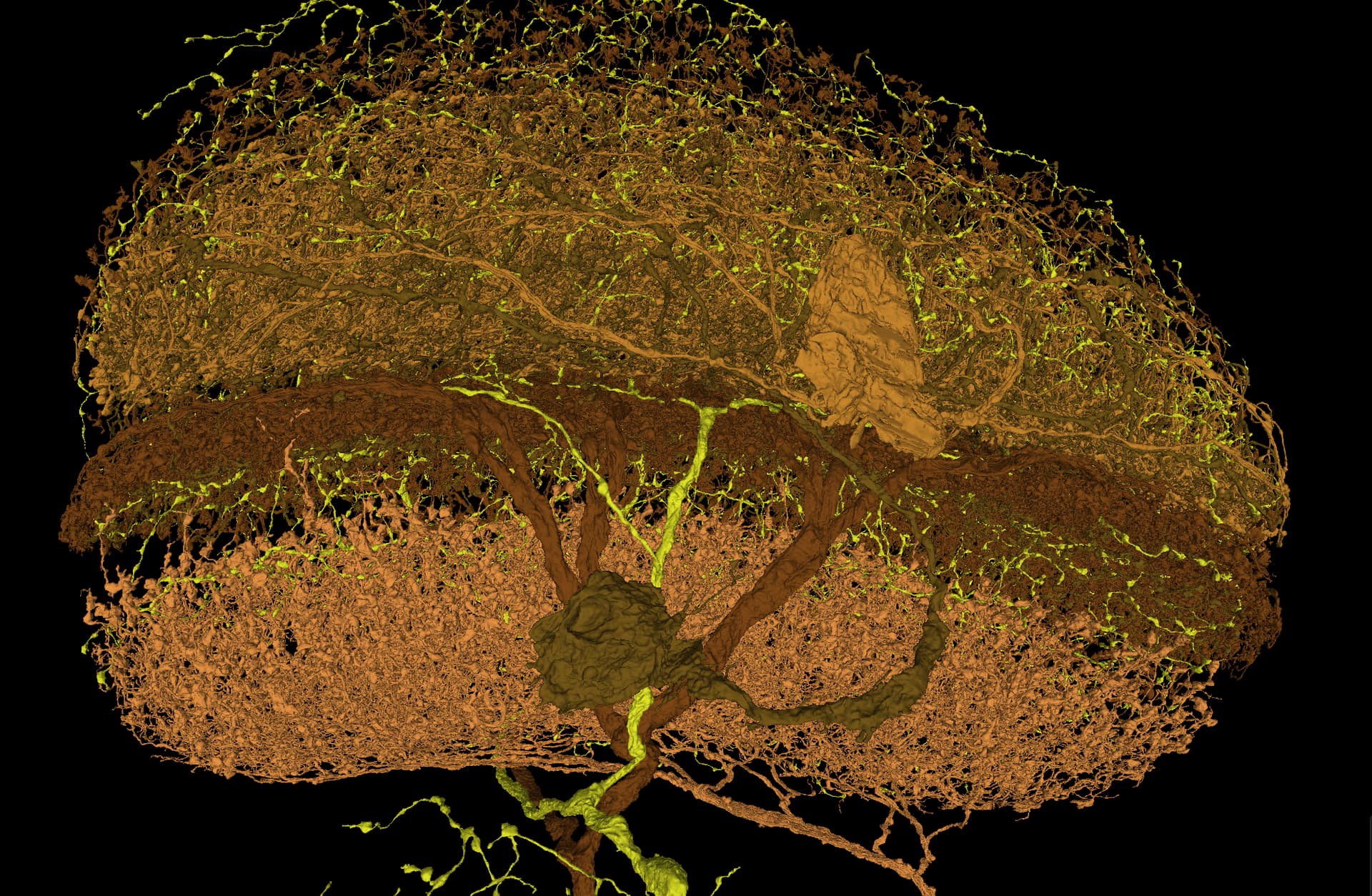
I wonder, what are those additional branches in these T cells (bottom left quarter of the pic). I’ve checked over 1000 of them and only these two have such extensions going around the Lobula.
https://ngl.flywire.ai/?json_url=https://globalv1.flywire-daf.com/nglstate/4808773340233728
Wowww this is amazing!!
Emil Kind has to this to say of these wild neurons: “I suspect some developmental variability. I also rarely see similar branching on other neurons. I could imagine that these are remnants of filopodia that are normally eliminated during development, but survived in this case.”