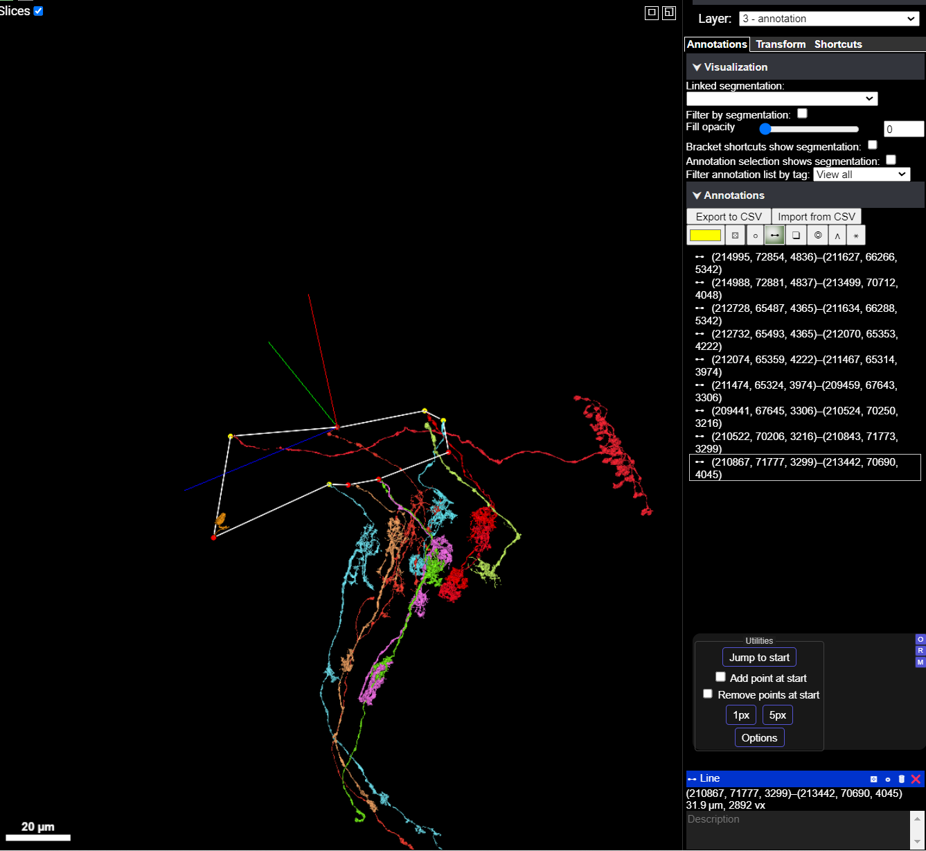
The annotation lines are just an indication, ofc i cant know if and how many more cells ‘outside’ of the annotated shape there might exist, so its more of a ‘current’ shape. I’m hoping that the more cells we are given to map the more cells we’ll find near/in/out of that area where the existing cells are which might give us a clearer understanding of the actual portion of the dataset that is within the ‘black spill’ area.
I immediately noticed something, while looking at the view/link you provided, that I had not noticed before. I haven’t heard it mentioned yet, so I’ll go ahead and point it out, just in case, in order to document it here in this thread.
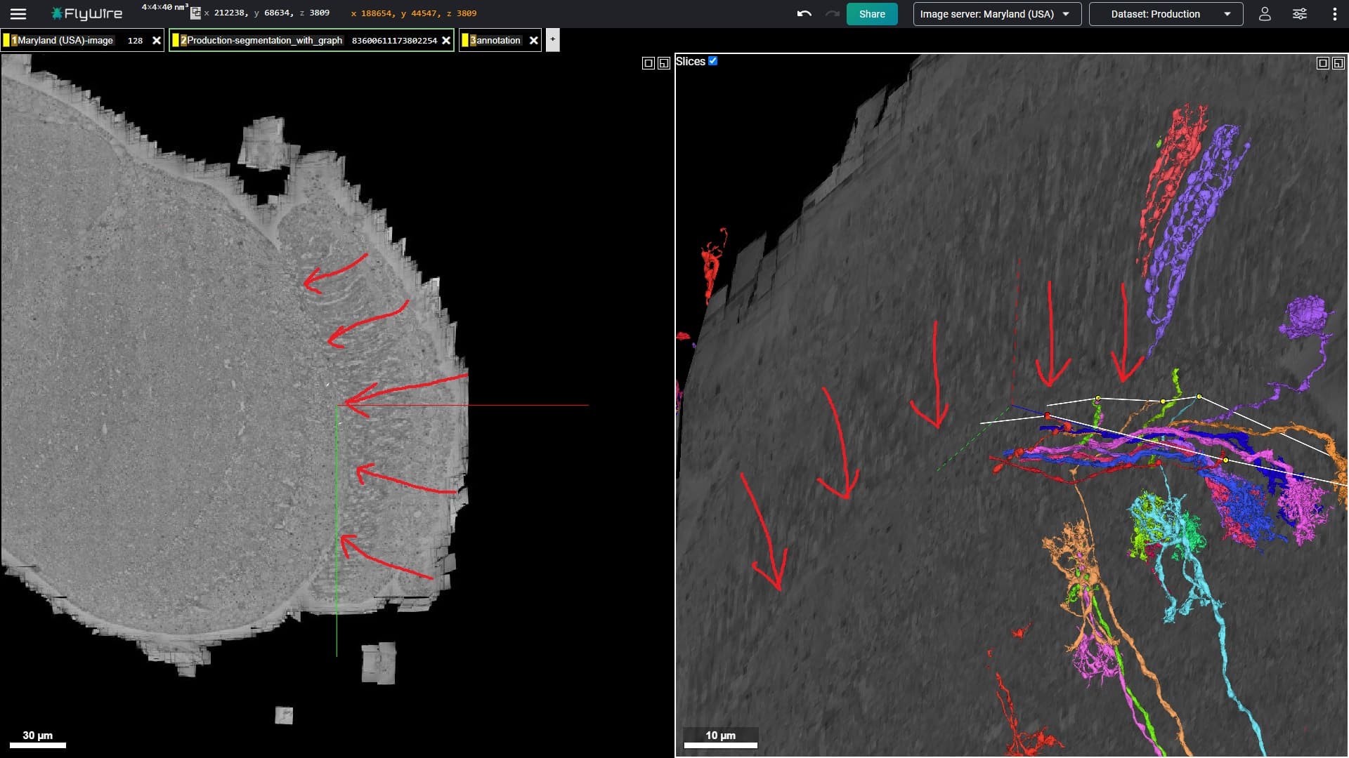
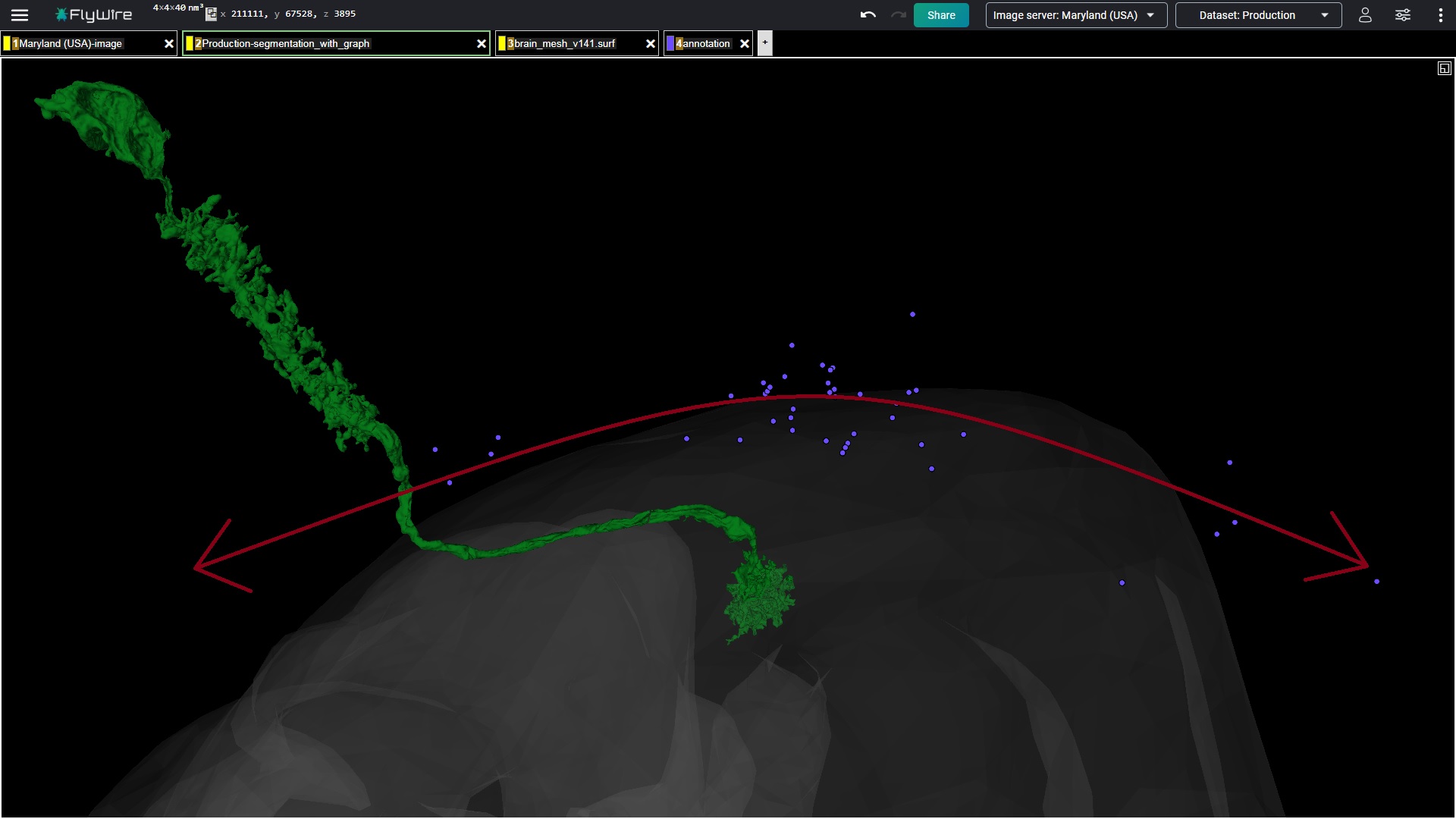
The surface roughly defined by your annotation points (in the view/link provided above) lines up almost perfectly with the natural boundary between the lamina and medulla (red arrows in the screen cap below…also note the T1 cells protruding through the EM, which form their characteristic “basket” structure in the lamina, after leaving the medulla).
I’m not going anywhere with this, at the moment…just thought it was worth pointing out and documenting here. Great idea, both of you, to form an organized approach on this. Cheers.
Amazing! Thanks for organizing this and adding a tab to the spreadsheet
Yes, this is the chiasm I’ve mentioned a few times on Twitch. It seems in our dataset, that there certainly is some difficulty segmenting/mapping the “first” chiasm that exists between the lamina and medulla. The tricky thing is that all the cells tend to criss-cross over each other making a big “X” as they travel from the lamina to the medulla. I think the diagonal crossing part may also be causing some issues with proofreading.
For those wanting more science, I briefly glanced over this 2019 paper, which does have a mention of the first chiasm (OCH1) in the Introduction and a few other spots. The paper primarily focuses on the second chiasm (OCH2) which exists between the medulla, lobula and lobula plate.
Thnx! And if im understanding this right (the paper) the chasm(s) comprise of a lot of axonal endings and few CBs? Maybe that’s the reason of the black spill area’s existence and/or origins? Maybe b/c of the increased synaptic activity between axons and whatever they synapse with the neuro-chemicals are very good at gathering up the dying agent which result(ed)s in black spill area?
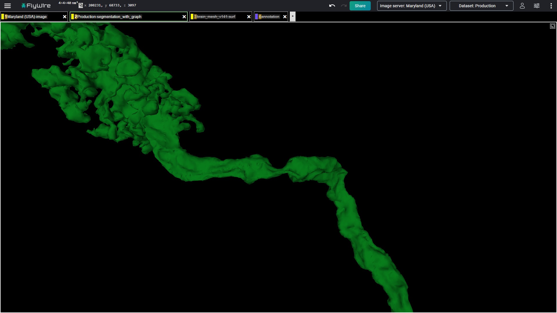
I can attest to how bad the AI has been at mapping axons coming from the retina:
these are merged retinal cells axons FlyWire im guessing that all the little ‘holes’ you can see scrolling up and down them are synapses with glia and/or other cells, the AI is HORRIBLE at these guys lol.
by comparison these are umerged and ‘finished’ axons: FlyWire
the purple one contains one of 2 key reasons why they are so heavily merged: white area between the axon and a another cell’s bouton, in this case if im not mistaken a LAWF cell’s axonal boutons. The other reason being the small ‘circles’ (non-scientifically named by yours truly haha) which im guessing are glial synapses.
@M_Sorek Thanks, M! Sorry, I probably heard you mention it, on Twitch, but did not recognize what you were communicating to us, at the time…just glad to have it documented here. And thanks for the link to the paper on chiasma!
@Nseraf Yeah, excellent hypothesis! As an additional consideration, there is a brief discussion in the slack forum about “damage” and “very dark tissue” in the lamina. Apparently, the lamina is easily damaged during dissection, which could also have contributed to what we see at the chiasm. Many of the T1 backbones that I have found look “pinched apart”, at some spots in the chiasm. I’m wondering if they were actually TORN apart, as a result of mechanical damage to the lamina, during dissection. This would also explain what looks to me like a river of mitochondria, at the chiasm (i.e. as the internal cytoplasm and organelles, of the torn neurites, poured out into the surrounding space)…or maybe the “river” I’m seeing is just glial structure…I’m still not sure what I’m looking at there, lol.
Not sure how to link a slack post here, so I’ll just call it out. The discussion is under “help_neuroscience”, on Thursday July 7th, and takes place between Qihua Chen and Sebastian Mauricio Molina Obando. Chen begins the thread with “Hi, I wonder what these areas…”.
Hey folks, just a small reminder that the Slack space for FlyWire is just for researchers. Not that everything is confidential, or that citizen scientists wouldn’t be hypothetically welcome under the right circumstances, but Slack charges per user so for budgetary reasons we have to currently restrict signups. FYI! Carry on.
Ooh thanks for letting us know, I wouldn’t have known about the cost per user issue on Slack. Shame we can’t have a read only version that doesn’t lead to additional cost (a fault of the Slack platform, I know!).
Oh, and it’s at least $6.67/month/user if paid yearly or at least $8/month/user if paid monthly.
If it’s possible and if it would help, you can delete my account from there (I don’t see a way to remove myself).
That said, I am wondering if we should try to isolate this data set as a specific focus, to garden cell bodies in the lamina as well so we can really work down the process of elimination.
Whatever the researchers need the most from us first of course.
Would there be any receptivity towards us self-funding our Slack account? Having a central chat that maintains history would be valuable, plus collabs on traces that could be threaded for responses w/ image attachments. I recognize there’s a certain duplication of what we have in combo of in-chat, annotations, and these forums, but having it all in one place would be nice.
Also I think Slack does free instances for non-profits up to 250 users.
As a possible resolution to the issue that has arisen, regarding community access to the Slack forum, perhaps the Citizen Scientist community could have a delegated Slack Representative? In other words, one particular “Flyer” with access to the Slack forum, who could inform the community of any relevant discussions taking place, and/or participate in discussions, on behalf of the community. In my experience, there is a lot of value in the discussions that take place in the Slack forum, and a community representative could provide access to those discussions, without having to bother the Admins every time we have a question about what is being discussed in Slack.
I am not suggesting that this the only way, or even the best way, to resolve this…but merely suggesting it as a possible compromise.
I came across this L2 cell that exhibits a highly localized and dramatic reduction in thickness/diameter, at the exact same location along the backbone as most of the “pinched” L cells that we have come across. So, I thought it might be worth comparing this “nearly pinched” neurite, with the current black spill point cloud (from the “black spill area” spreadsheet). And sure enough, it lines up perfectly with the current point cloud, so…the apparent damage to this “nearly pinched” neurite may provide some insight into what has happened to the other cells in the “Black Spill” area.
And thanks again to @Nseraf and @annkri for taking the initiative to map these discontinuities, and identify the affected area (see point cloud in the link below).