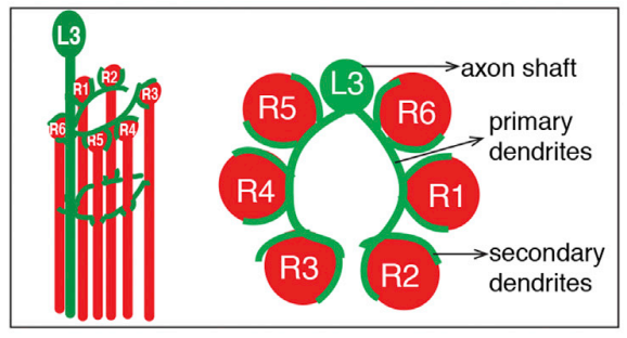
This paper suggest, that one can also differentiate the R1 - R6 neurons, by finding the L3 neurons and then, going clockwise, there should be R6, R1, R2, R3, R4, R5.
Looking at other images (including some in the “Visual…” thread on our forum), I’m quite sure, that the last image is a mistake and that it should be possible to identify all R subtypes by their position in relation to the L3 neuron.
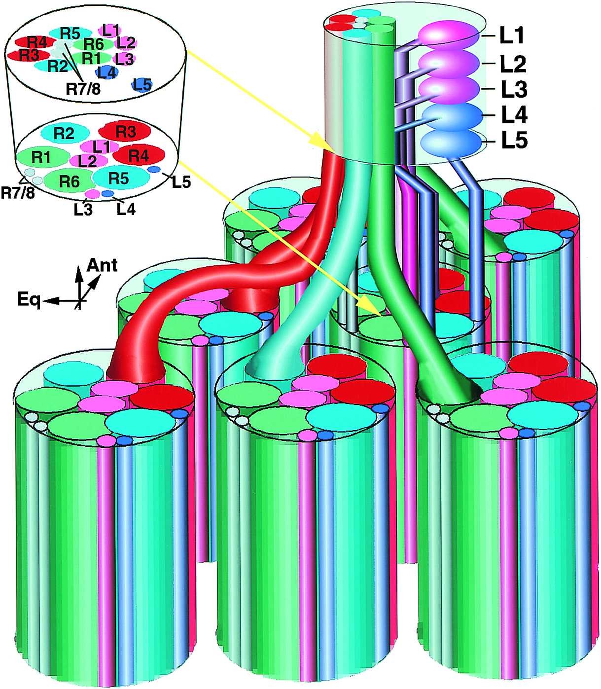
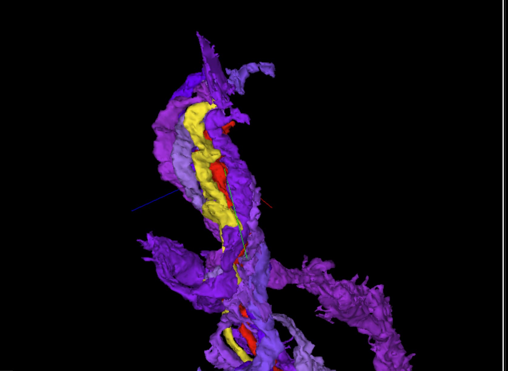
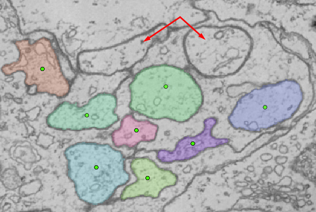
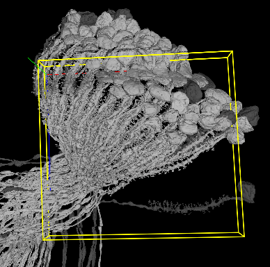
In the top of our cluster it looks like R7 (yellow) is outside in the ring with R1-6 while R8 (Red) is more hidden in the middle. Have only cheecked a few and will have to check that this is always true and in betwheen R1 and R6 as suggested in the illustration before using that as a way to identify the rest. But this might be a easy way to identify all of the R - cells if true (also DRA 7/8 and R7/8 that is cut of by the gap.)
I believe, the top part on your picture is still in the retina. We call look there too, but it should be easier to look into the bottom part - in the lamina. And here all the six outside neurons go into separate cartridges (the last picture in my previous post shows the transition).
So, to identify the R1-R6 neurons, one would have to find all the other R cells from these new cartridges, then all the L3 cells for them and only then try to recognize the order.
In other words, currently we have 6 R cells in the link, but we need 36 + 6 L3 to identify everything.
i agree that we have to use the description in published papers too start the identification prosess either using L3 or connected partners or even both. But if it is as i think that R7 is part of the circle in the retina and the other cells either don´t change place or change place in a regular manner. It would probably be a much easier metod to use. Both since you would not need any extra cells and since they are in easy identifiable clusters. from you picture it looks like they are in the same order in retina and lamina, but the clusters in lamina is turned 180 degrees compared to retina and have more other cells around it, and in my experience it is much harder too se the different clusters.
Oh, right, ok. I thought, that the usual case, is that the retinal part isn’t available for most of the cells, hence the suggestion to identify them by the lamina cartridges. However, if those part are visible in the dataset, then indeed, that should be much easier to go this way and only use the lamina identification as a backup for some edge cases.
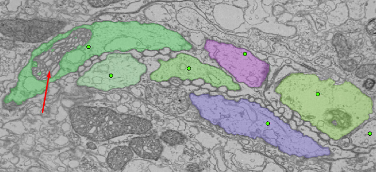
Other annotation layers remain on this if you want to view them - “L/T” represents any found lamina monopolar or T cells found, while “Non-PR” is just a jumbled mess of glia, Lai, etc - basically my visual “exclusion” notes. All on (green = PR, blue = L/T, red = extras):
The edge of the data can be tricky - these are both blending in a bit with the surrounding tissue. One of them gave away its position with the cell structure.
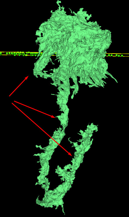
As for 3D, just one tip on what to look for in glial masses are structures that look like these:
so there is either two R1-6 that you have not found, our that is right outside your sector, could ofc be that there are several clusters on the border beeing splitted also.
How strict to the zones should we remain for this? Not just in terms of inter-zone overlap, but also for areas not covered by a zone at all (if there are any). As a bad example:
I’m assuming these low ones should still count as part of my zone (obv will still id them even if not), but much further to the right and it exits the search area completely. If there are any out there, are they considered lower priority atm, or equal prio and the zone just doesnt cover them because its a curve?
I guess ultimately my question is: should i only care about cells directly within the bounding box, or should the zone invisibly extend in z (and x also?) direction too?
As you said, it’s a case of the areas are all “equal prio and the zone just doesn’t cover them because its a curve”. So we’re approaching the quest to map the photoreceptors from two sides, (1) trying to map all the cells within a zone and (2) by searching for each column via the Mi1 and L1 cells. There are some tracers working in zones in the Z direction in a layer below the current Flyer zones and there are some doing the second Mi1/L1 method.
It’s okay if you want to stick to only mapping the photoreceptors in that one area/level as outlined by the bounding box - or - if you keep finding neighbor photoreceptor friends and go a bit beyond the lines, that’s totally cool too! If you’ve seen me on Twitch, I tend towards the latter and go out of bounds here and there (I get easily distracted with finding friend cells )
Overall, I’d say for our goals as a group as a whole it’s best to prioritize getting everything within the Zone first, so we can keep track of progress and know that we have covered that area. Beyond that, feel free to fly around!
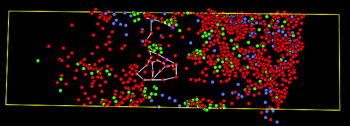
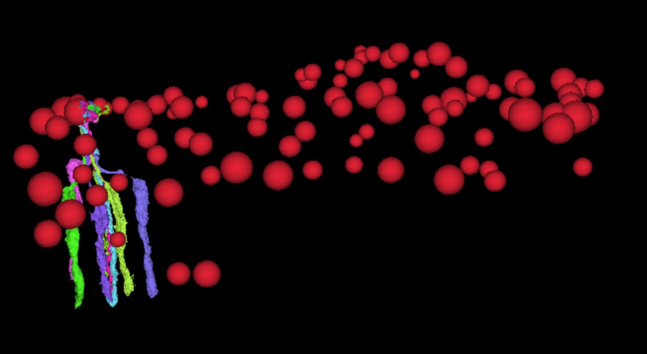
My first 100 complete clusters of identified photoreseptors with a total of 800 cells. Each cluster is numbered with a corresponding group of id´s in this sheet cell farms - Google Sheets
Have found every L1 that passes below zone 13, estimating around 50 or so (they curve too much for a count to be too accurate ). A fair bit of overlap occurs with zone 12, so who knows ¯\_(ツ)_/¯
Most of these ones dont pass through the official zone boundaries, but i expect the ratio that do will be quite a bit higher for the “inner” zones
Found an interesting R1-6 interaction, thought I’d share and maybe get some scientist feedback?
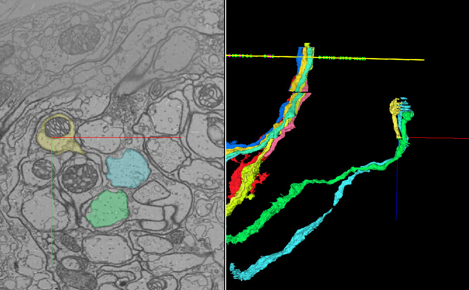
While I’m used to seeing R1-6s ‘interlock’ with each other, this particular junction looks like fusion in the 2D section. There’s more than just a passing portion of the cell from one to the other: the edges genuinely seem to conjoin for a few 2D slides afterward.
Link (this is before I finished editing either cell): FlyWire
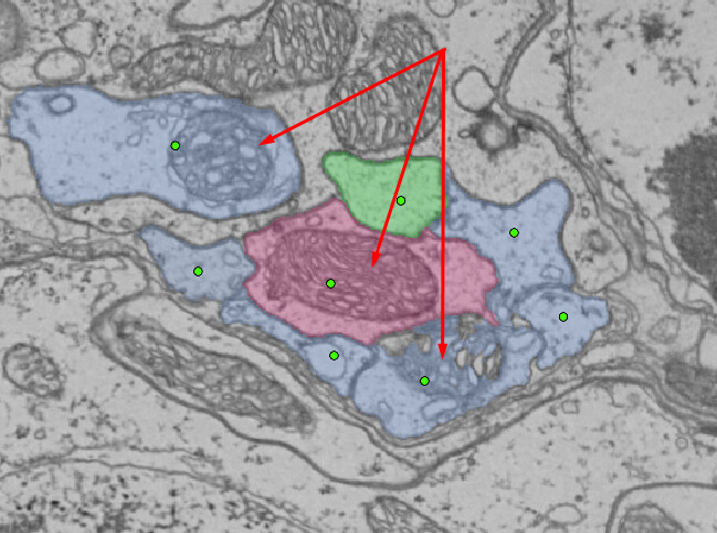
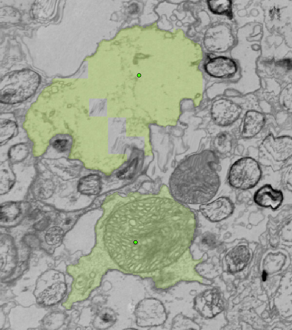
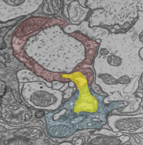
Here you can see the passing of cell matter between the two cells (highlighted yellow)
Here you can see that the cell barriers do seem to conjoin as they pass this material:
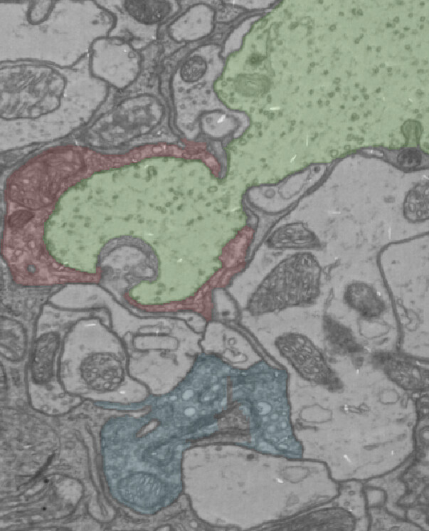
It could just be some rough borders, given the area, but I’ve never quite seen an interchange like this one - usually there is a very clear delineation between the ‘interlocking’ parts (such as we see with this neighbor):
Very cool! I think it is most likely that the barrier broke down or got overstained here. Especially since the interlocking projection ends in the lower blue neuron.