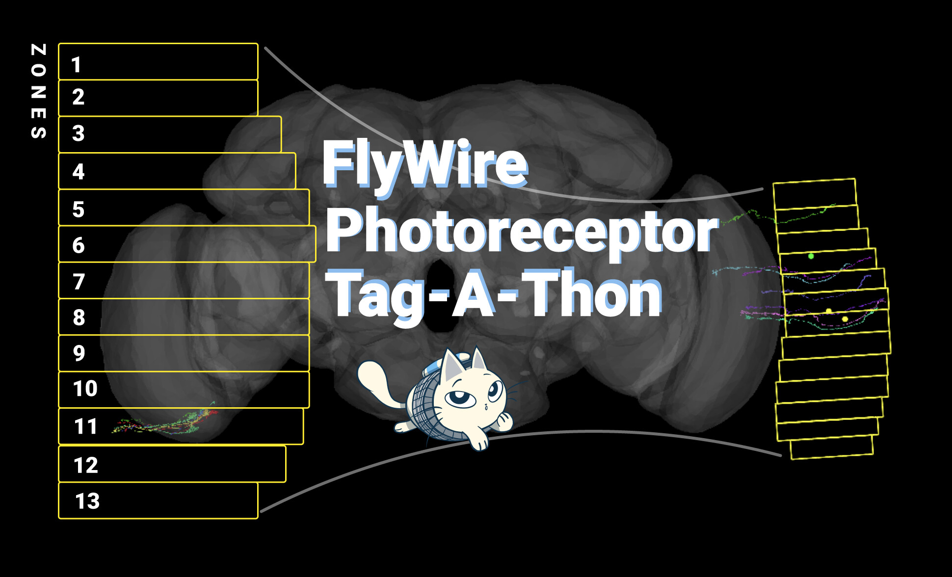
We’re back with another round of Known Unknown aka cells that we know are there but that aren’t tagged yet in the connectome. This round is a critical step in the sensory pathway of vision: photoreceptors. Will you help us find and tag them?
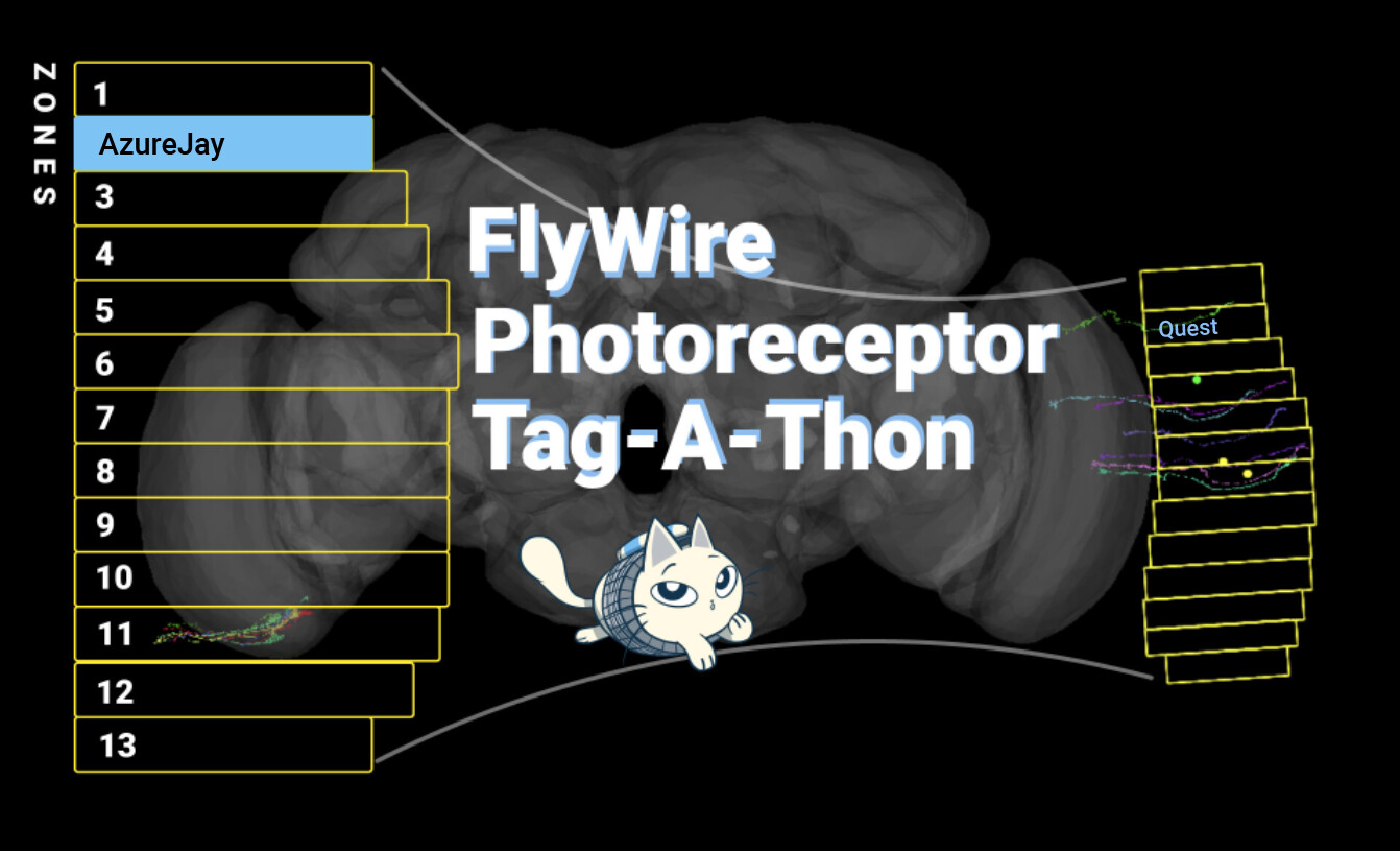
Update 7 March: the first sector has been completed by AzureJay!
Update 25 May: As part of a joint effort from Flyers, Princeton lab members and researchers, the Photoreceptor Zone Quest in the right optic lobe has been completed!
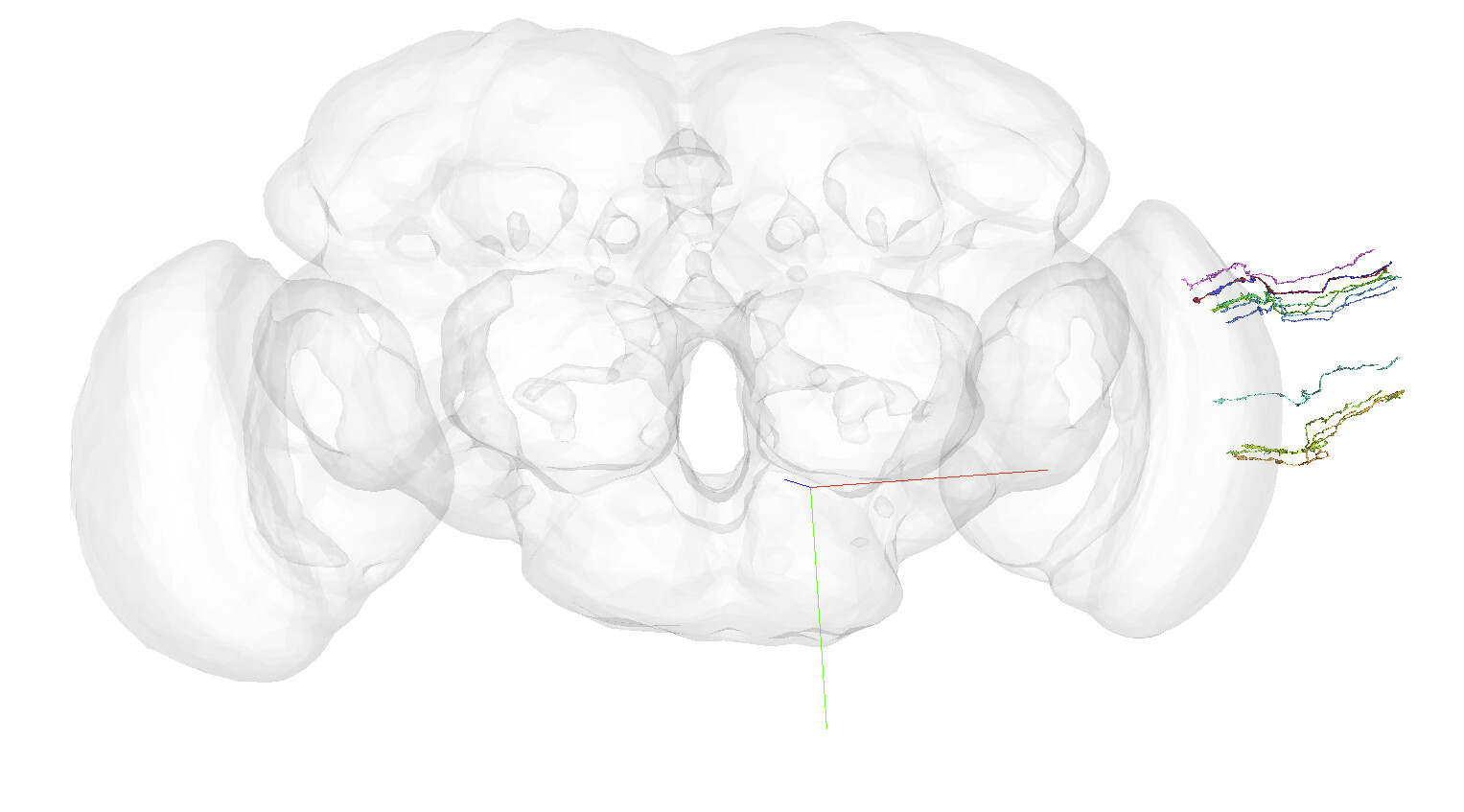
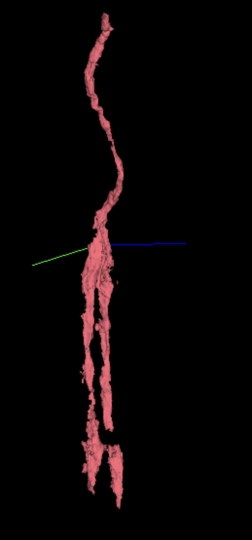
Example R8 photoreceptors. There are only 11 tagged in codex → FlyWire
Nested dataset update →
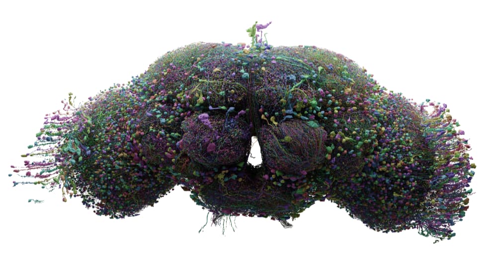
Snapshot of ~7,000 neurons from Sven. We can’t seem to render more but interestingly even this few cells starts to look densely filled in. Brains are so complex it is nuts! Please don’t share this image outside the FlyWire forum as it is a work in progress for one of the figures in the upcoming preprint paper.
i am working on them and have 90 R7/R8 FlyWire and 403 R1-6 FlyWire with more on the way, but it goes rather slowly to find them since i need to free them from the glia cells and each other
i am working in the right lobe and there few of the photoreceptors i find are proofread, ofc the way i am looking (finding glial segments and freeing the the R- cells from them i will mostly find cells not proofread already, but i am also trying to find the cells missing from the clusters of 6 (8) R -cells)
i know Nik did some proffreading of R - cells in the left lobe, but not sure how much that was.
Since many of the R- cells are bellow 100 synapses or difficult to spot in the glial i am not sure how much effort have been used trying to finish them of
Nice find! We were just looking at finding an example of this in FlyWire. Any chance you’ve already found one - that is, all the photoreceptors of one eye cartridge?
Ooh, great project! I’ve been out of commission for the past month, and have a project I need to finish up first (not too far from completing) and then I’ll jump in and claim a sector.
Are we only loosely ID’ing R1-6 for now or are we trying to specifically identify each one?
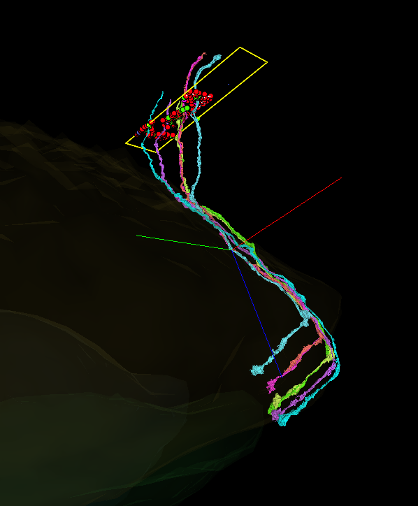
If so, am I right that the ones with a longer cluster of synapse points are R8 DRA, while the ones that have just the bottom cluster focus are R7 DRA? EX:
i think the two green, purple and blue to the right of your picture are DRA cells the rest are normal R7 /R8 since you can see one short and one long + it is not on the edge
i am guessing the light green one is DRA 7 and the other DRA 8, but you will have too cheek the connections too be sure.
R7 DRA is the only one that do not have connections to TM or TmY cells (particularly Tm5 and Tm 20 )
Yup, annkri is correct. The easiest way to identify R7/8 and the DRAs is by grouping them together in pairs. You can compare them by looking at what one is shorter than the other and also where they are having their axonal terminals.
DRA R7/R8s have their axonal terminals bunched together with each other (typically the shorter of the two is the R8). As annkri, said that is most likely because the DRA R7 doesn’t have Tm connections so instead it is terminating with its R8 partner.
For “normal” R7/8s, you can identify them by the fact that the R8 cell is much shorter and terminates “higher” up than its partner R7 cell. This is a quick way to determine if the cells are not the DRA type.
I’ve added tabs with each of the labels for the cells in your example here: FlyWire
(I also added the partners for some of the cells in your example since it’s much easier to identify them by pair).
PS. To help remember that the R8s are the shorter of the two… “R7s have projection, R8s truncate” (Special thanks to @Celia_D for helping come up with that little saying)
PSS. Here’s another example of a DRA R7/R8 pair just in case: FlyWire