i added a link too the post about both Dm9 and L cells (first in discussion) in the sheet so it is easier to find when needed
3 Likes
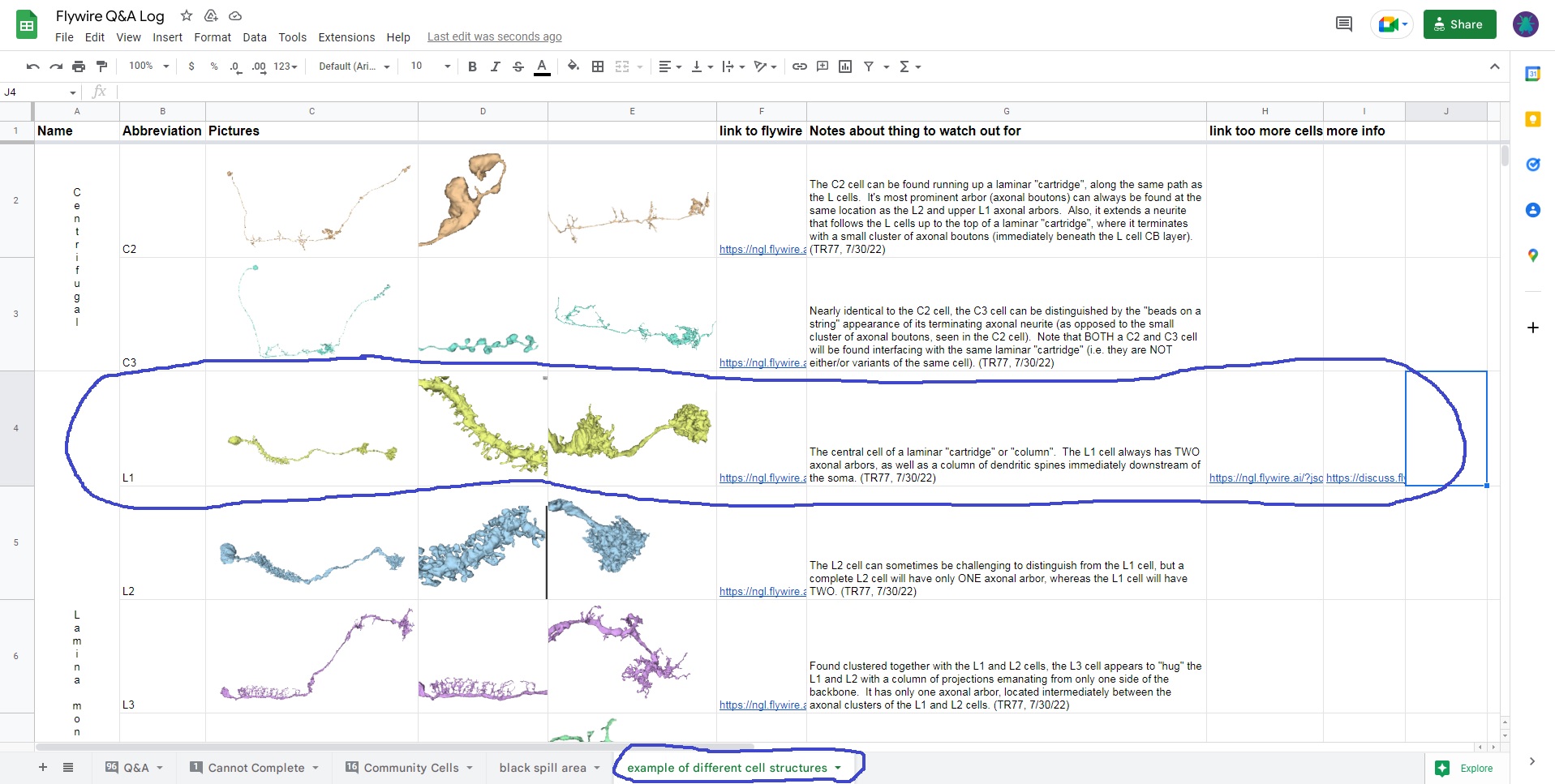
Okay, so…check out how awesome this is!
In the L1 cell entry, in @annkri 's “example of different cell structures” spreadsheet, you can now view community selected preview images (columns C thru E), view an officially documented example of the cell in our dataset (column F), read a brief community description of the cell (column G), view a link containing several community identified examples in our dataset (column H), and even click a link to a discussion about the cell in the community discussion board (column I)!
My goal now is to complete an entry like this for every cell type in the optic lobe…b/c that would just be fantastic, lol.
Cheers, all.
6 Likes
Medulla Intrinsic 1 (Mi1)
The Mi1 cell features a single branch with three total clusters - two dendritic, and one boutonic. The soma begins outside the medulla, and the branch takes a hard angle turn (approximately 90 degrees) into the medulla.
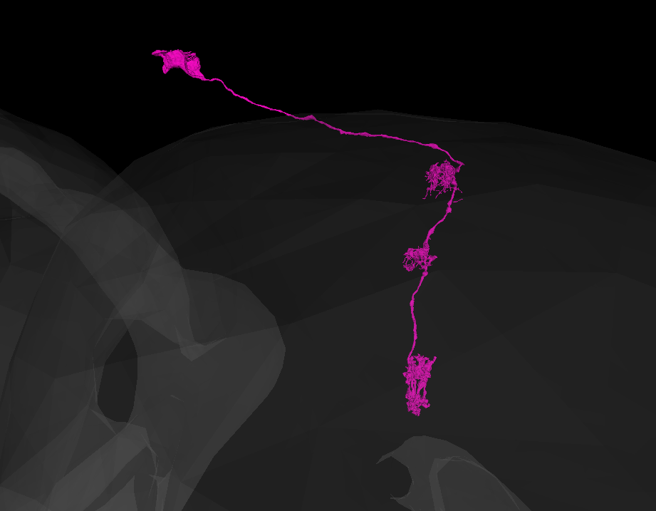
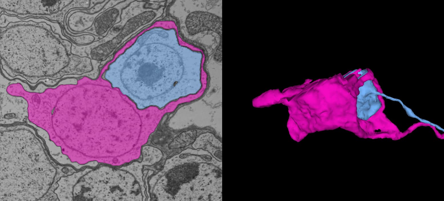
The soma has a ‘blanket’ layer which wraps around another cell’s soma (a TM type):
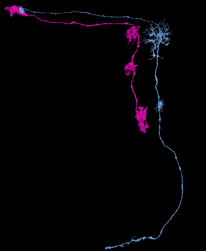
The first two “dendritic” clusters are roughly spherical. They wrap around and interface with an L1 cell.
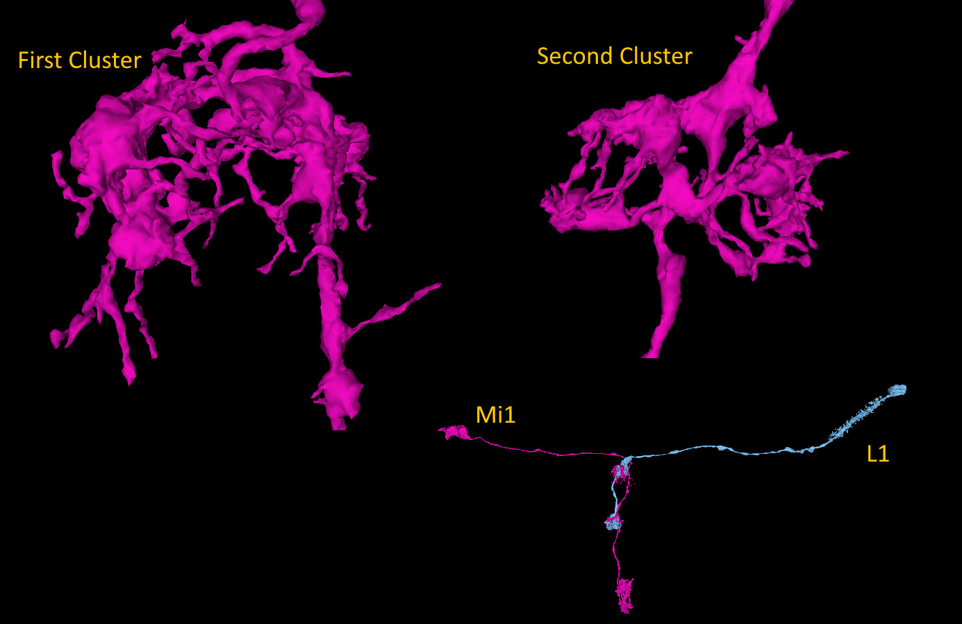
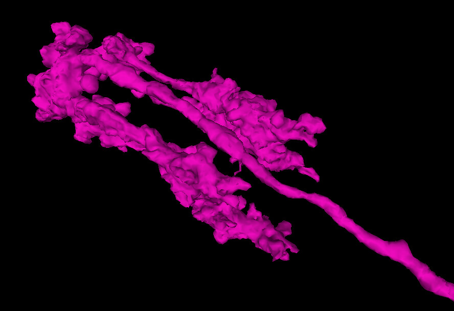
The final “boutonic” cluster features several (3-4 seems to be common) bleb-type bundles that go upward along the axon:
Known examples: FlyWire
5 Likes
Avesome post AJ
After working on the sheet, i see that the part i have had too little focus on is where in the brain you can find the cell. Partly because i did not see the importance of this at the beginning and have just continued using the same pattern for the pictures. And partly because it is very difficult to get good pictures showing this when it have to be visible on small pictures.
With a forum post i am thinking this would be a good place too show this,
maybe a short video zooming in/out and around the brain mesh might be a good way? maybe even use the coloured brain parts that amy have showed us, if that would show it better.
5 Likes
I’d just like to add, that the type of the structure seen on the last picture is sometimes called bleb-type arborization.
3 Likes
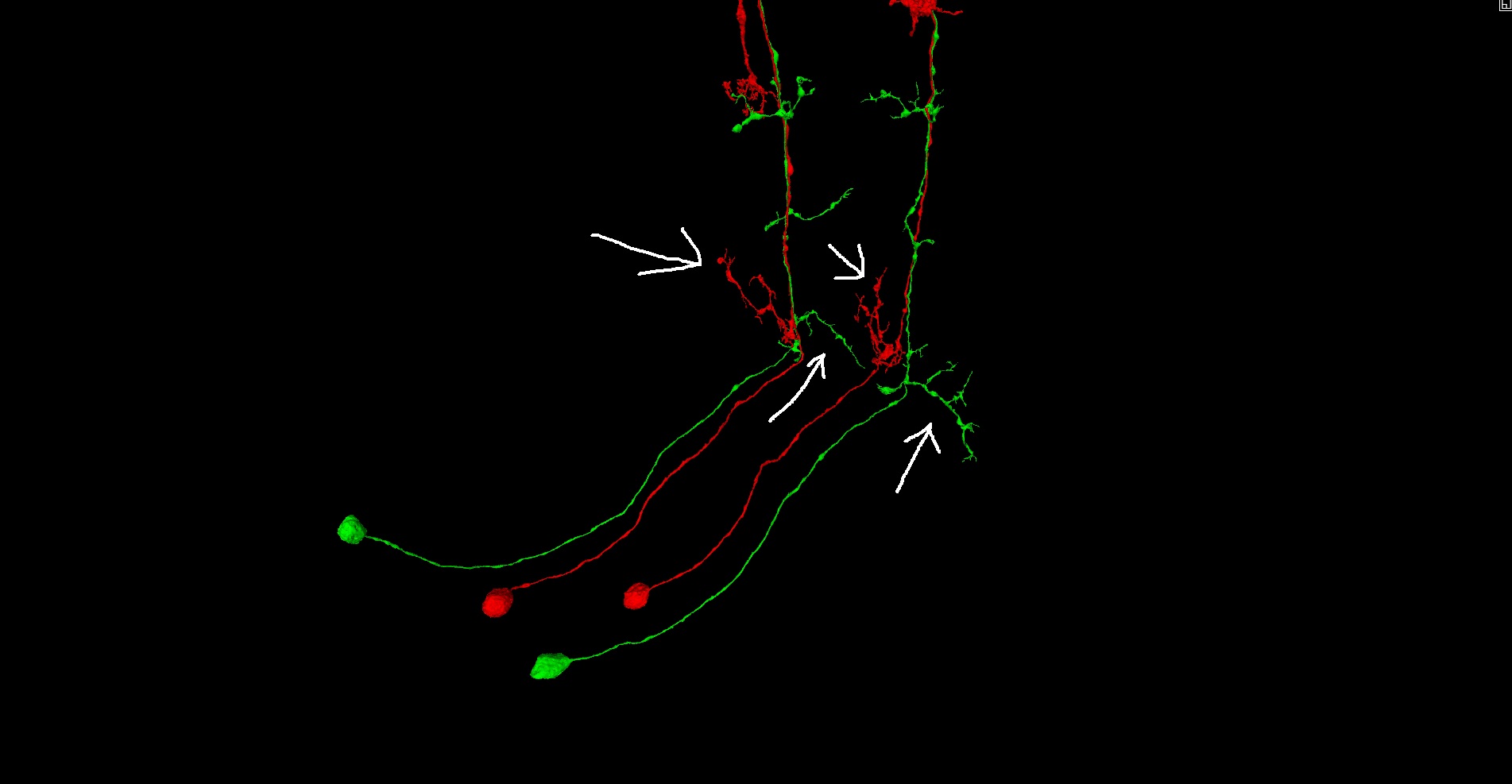
Miscellaneous useful structural pattern…
Noting here, a pattern that I have been seeing, in the arborization of Centrifugal C2 (green) & C3 (red) cells, at the location of the 90 degree bend. This pattern can be useful in identifying and reassembling fragmented segments of these cells. In other words, when you locate a soma with a piece of the backbone attached, you can often quickly determine which cell’s soma you have located, based on the pattern of arborization at the 90 degree bend.
https://ngl.flywire.ai/?json_url=https://globalv1.flywire-daf.com/nglstate/6193692196995072
4 Likes
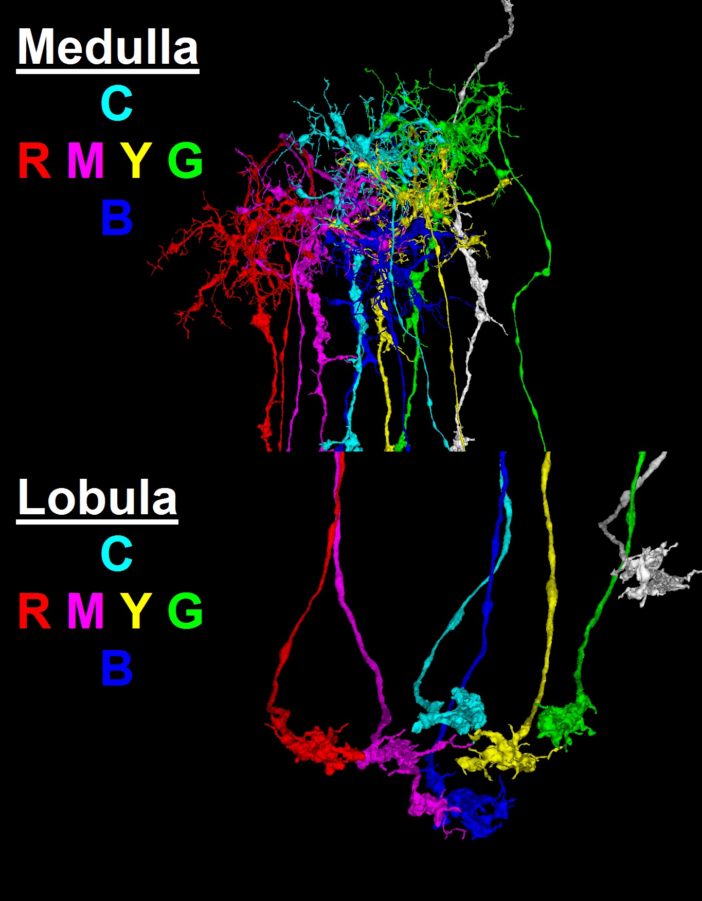
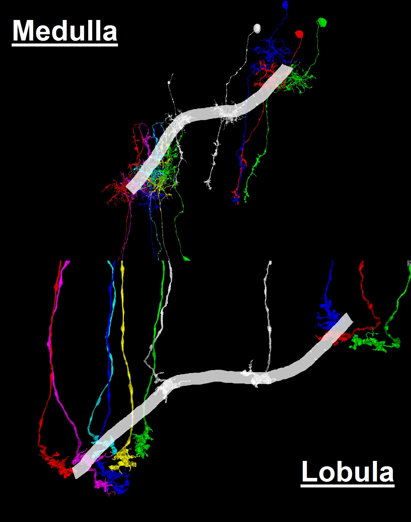
Miscellaneous useful structural pattern…
Noting here (or rather confirming here), that the relative arrangement of Transmedullary Tm/TmY cells in the medulla, is preserved in the lobula. Specifically, in the diagrams below, the arrangement of the dendritic arbors of Tm4 cells, in the medulla, are shown in comparison with the location of their axonal arbors, in the lobula. As can be seen, the relative arrangement of the cells is preserved both locally, and over large areas as well. I have not verified this pattern for every single one of the many types of Tm/TmY cells yet, but have observed it in several already, and felt it was worth pointing out and confirming here.
This can be useful in locating and reconnecting separated pieces of these cells, in two different neuropils. Basically, if you know the location of the cell in one neuropil, then you already know the location of the rest of the cell, in the other neuropil.
As an aside, it is perhaps not surprising, in some ways, that the relative arrangement of the cells would be preserved in this way. The Tm/TmY cells, after all, are presumably transferring the entire visual field from the medulla, down to the lobula. So, it is perhaps of little wonder, that their relative “pixel locations” (if you will) are preserved in the transfer.
https://ngl.flywire.ai/?json_url=https://globalv1.flywire-daf.com/nglstate/5450687148195840

5 Likes
Transmedullary 2 (Tm2)
The Tm2 cell has a straightforward branch pattern with three total clusters - a dendritic arbor, and two small bleb-like clusters. It starts above the medulla near the edge of the EM dataset, passing through into the lobula along the corridor between the lobular plate and lobula.
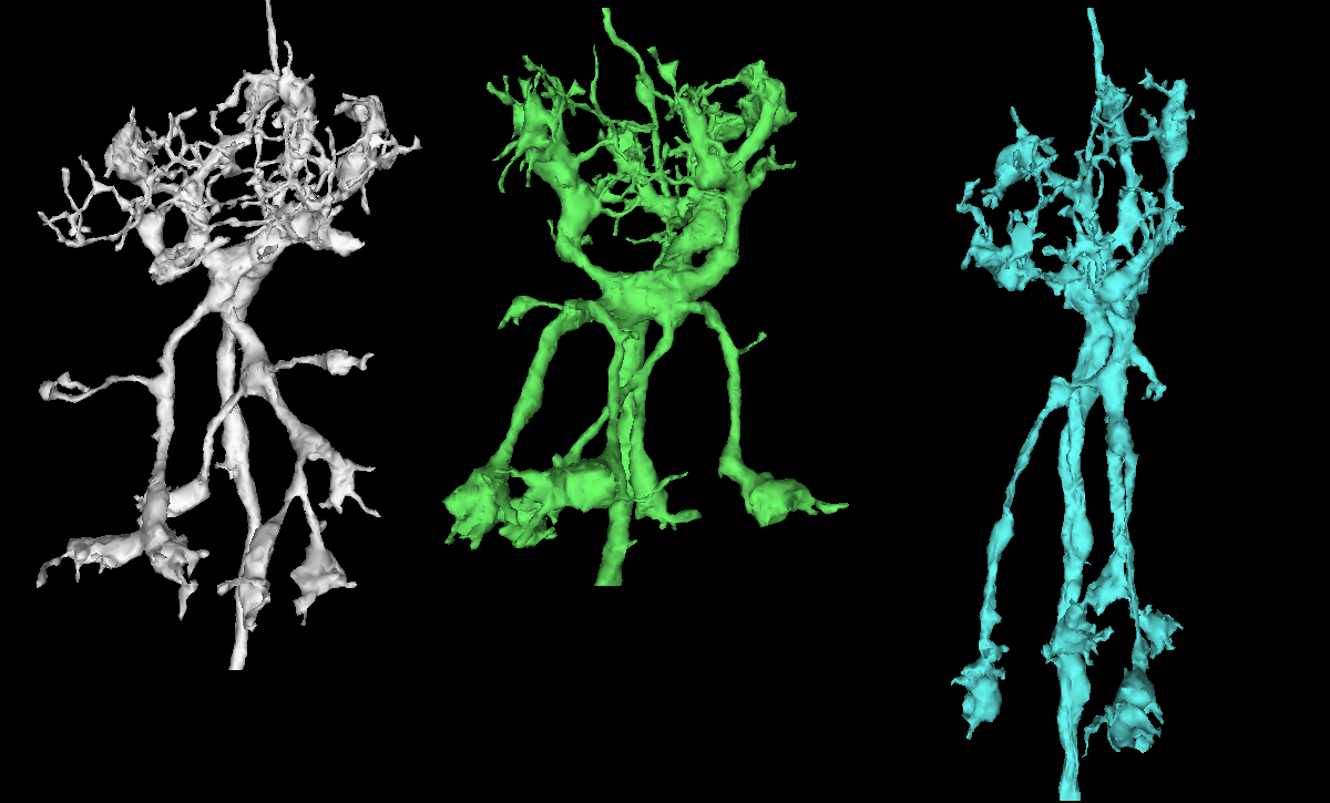
The first cluster is an arbor found in the medulla and has a unique “X” type structure, with the top of the X having small dendritic twigs and the bottom having larger bouton extensions. This is the most recognizable part of the cell.
The second cluster, in the lower medulla, features only a couple bleb-like extensions.

The final cluster, found in the lobula, has only one bleb-like structure.
6 Likes
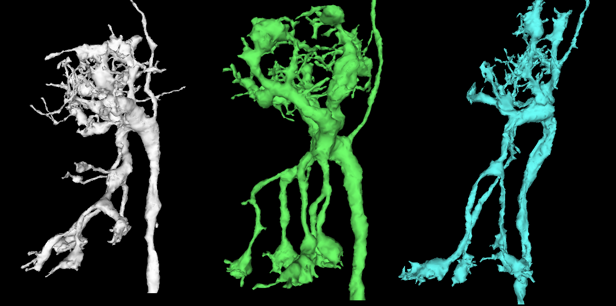
@AzureJay “Great minds…”, AJ…I’ve been working on Tm cells lately as well. ![]()
Also, note that as per my previous post on the preserved arrangement of Tm/TmY cells in the medulla vs lobula, you can see from the last two graphics in your post that the arrangement of the Tm2’s is preserved as well. This is also apparent in the link you provided. So, this example demonstrates that the preservation of arrangement goes for Tm2’s as well as for Tm4’s (and for many others, in my experience).
Outstanding work, AJ! ![]()
![]()
2 Likes
Created a small table to show differences between TmY5 and TmY5a, which are hardest to differentiate for me. The table describes arborizations in different layers of optic lobule’s neuropils (according to FlyBase.org).
The easiest ones are in the Medulla layer - TmY5 starts its arborization “lower” and it’s usually finer (no bleb until M10).
| Layer | TmY5 | TmY5a |
|---|---|---|
| Medulla | ||
| M1 | ||
| M2 | fine | |
| M3 | fine | fine |
| M4 | fine | |
| M5 | fine | fine |
| M6 | fine | fine & bleb |
| M7 | fine | |
| M8 | fine | fine & bleb |
| M9 | fine | fine |
| M10 | fine & bleb | |
| Lobula | ||
| L1 | ||
| L2 | ||
| L3 | ||
| L4 | fine | fine & bleb |
| L5 | fine & bleb | fine & bleb |
| L6 | fine & bleb | fine & bleb |
| Lobula Plate | ||
| LP1 | fine & bleb | fine |
| LP2 | fine & bleb | |
| LP3 | fine & bleb | fine & bleb |
| LP4 | fine & bleb |
5 Likes
This is a breakdown of the medulla layers (M1-M10) as described by The Fischbach Paper. This is useful in knowing how these layers are defined, because they also help us, in turn, know how to define some of our neurons.
Note: I use “inner” instead of “proximal” and “outer” instead of “distal” for reading clarity’s sake. Both terms refer to position to the central brain (inner/proximal = against central brain; outer/distal = away from central brain)
| Medulla Layer | Defining Characteristics |
|---|---|
| M1 | Extent of L1 Arborization in the outer medulla |
| M2 | Extent of L2 Arborization. Contains outer L4 arborization, T1 arborization. Inner M2 defined by outer expansion of L5 (which fills M1 and M2). |
| M3 | Extends from inner border of L2 arborization to L3 terminal. Contains main medulla arbor of Lawf, most R8 terminals. Border between M2 and M3 marked by Dm3 |
| M4 | Slim layer between L3 terminal and inner L1 arbor. Contains inner L4 arbors, and long R8 terminals. |
| M5 | Inner L1 arborization. Contains inner L5 terminal. |
| M6 | Between L1 arborization and serpentine layer. Contains terminal enlargements of R7. Can be subdivided into M6a and M6b depending on R7 terminal. |
| M7 | Serpentine layer. Axons of many medulla tangential (Mt) neurons. |
| M8 | Stratified arbors of some Medulla intrinsic (Mi) neurons, ex Mi3-Mi7, Tm and TmY neurons, and C2’s lower arbor (M8-M10). |
| M9 | Houses several types of Proximal Medulla (Pm) cells. |
| M10 | Height of the bushes of T4 neurons. |
4 Likes
@Krzysztof_Kruk @AzureJay Nice work, KK and AJ! ![]()
On moving forward, with Tm/TmY cells…
My primary goal is, of course, to proofread and complete the cells of the optic lobe. As such, my primary interest in cell type classifications is to aid in that goal (i.e. if you know what type of cell it is, then you have a pretty good idea of what the cell “ought” to look like, when complete). At some point though, assigning cells to further and further subdivided categories becomes a purely academic exercise, from the standpoint of proofreading. As an example, consider the distinction between the Tm1 cell, and its Tm1a variant. In my experience, the examples of Tm1a that I have found, have always been in place of the Tm1 cell, in a particular medulla column, rather than being an additional cell in the column. From the standpoint of proofreading, and useful structural patterns to aid in proofreading, it makes no difference to me whether it is a typical Tm1, or some slight variation of the Tm1 theme…it is sufficient for proofreading purposes to simply know that every medulla column has a Tm1-ish cell, with a proximal medulla arbor somewhere near the distal end of the Mi1 axonal arbor, and an axon extending somewhat superficially into the lobula, etc.
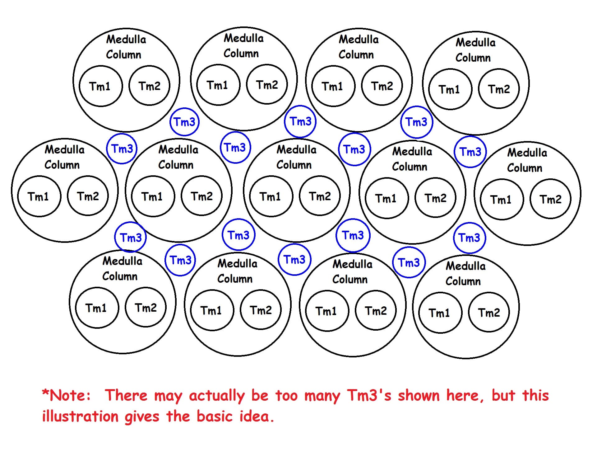
With the above in mind, I would personally like to organzie the Transmedullary cells into two broad categories - those that are coaxial with the medulla columns, and those that are interstitial to the medulla columns. The figure below illustrates what I mean by this distinction. In my experience, Tm1 and Tm2 can always be found within, and coaxial with, a medulla column. The Tm3 cell, on the other hand, can always be found in the interstitial space between medulla columns. I would then like to come up with a count of all coaxial Tm/TmY cells, in a typical medulla column, and a count of all interstitial Tm/TmY cells, at a typical interstitial location…and then compare those totals to the Fischbach classification scheme. Why? Because Fischbach acknowledges in the paper that, for example, Tm8 and Tm22 may actually just be slight variations of the same cell type, and my experience suggests that Tm5 may also be yet another variation of that same cell type. There are other examples, but this paragraph provides the basic idea of what I am looking for.
I am not asking the community to do anything here, I will pursue this avenue of exploration myself (although anyone is more than welcome to help, if they like)…just providing an update on where I am at, regarding the question of identifying Tm/TmY cells.
Cheers, all.
6 Likes
How to tell apart ‘normal’ cell Somas/CBs from Glia ones?
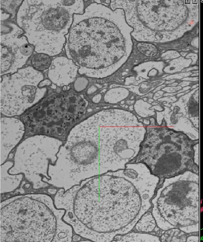
Iit’s not 100%, but very often the glia ones are going to be nucleus and surrounding area all ‘pitch’ black, unlike the non-glia neuron CBs which are ‘white’ (or shades of grey), like in the picture, the 2 glia cells are much blacker than the other cells.
The other way to know is they’re usually larger than the average cell, with the exceptions of the really large non-glia cells’ CBs.
FlyWire the link contains 3 black CB glia cells and one large CB, the 3 are not reconstructed so they’re probs mergery.
5 Likes
Yes, and often the branches themselves are usually at least a little bit darker for glia cells.
Maybe the matter, from which the cells are built is denser and keeps more osmium (or whatever was used for staining), that the neurons.
4 Likes
Hi all,
Something has come up on my end, so I will be away (afk) for a while. Keep up the good work, all!
Cheers.
5 Likes
have anyone testet/noticed if the widefield amacrine neurons make layers thight layers like the dm 4 does? Have a feeling that if it does that might help finding the missing clusters
5 Likes
Didn’t test, but that’s a good idea.
2 Likes
Idk abt a specific cell type but in the CBs layer in 182648, 80934, 5572
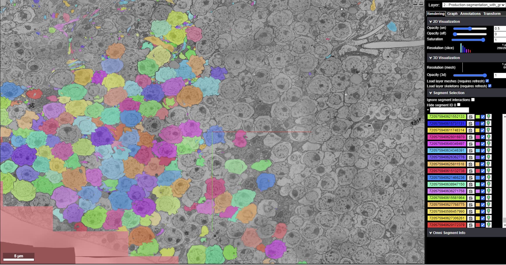
and the one ‘above’ I have noticed that the stems/backbones going from the CBs to the dendrites/axons etc all cluster around like this;
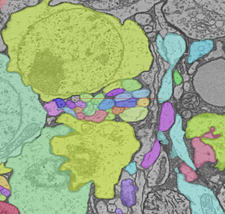
which isnt 100% that the CBs will be in one place but they look like this:
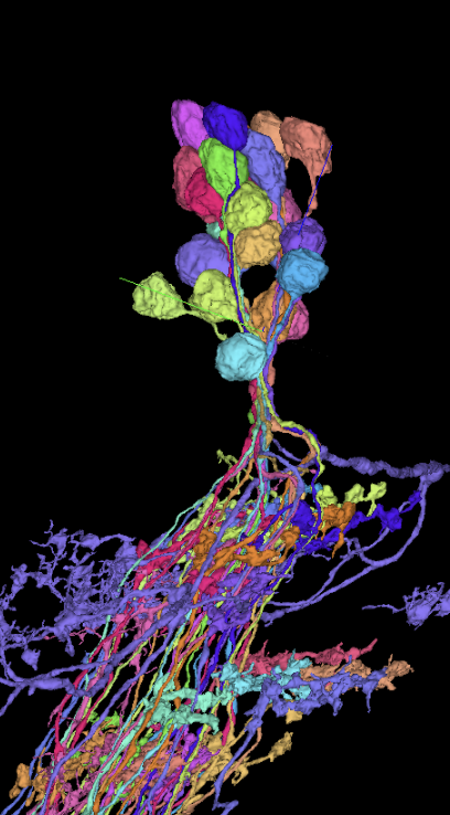
So, I’m thinking this: Say that u have either a CB w/o backbone, or vice versa backbone w/o a CB, scroll a bit up or down and find the other backones in the ‘bunch’ (or bucket? following the DMs flowers analogy lol) and if u have say 10 CBs/Backbones and 8 or 9 of them have a match in the boucket/bunch then it becomes a whole lot easier to find the missing backbone/CB from 1 or 2 than 1000k (cells in the brain lol).
Disclaimer: ive not actually tried to find a backone’s CB yet but I keep seeing layers of ‘endless’ CBs and the CBs seem to alway seem or tend to go to clusters of backbones. May not end up helping - this observation- as the black spill area might just cover up/destroy the pathways from cb to backbone, but maybe if some of the other cbs’ pathways in the bunch are restructurable/ or restructured then deductiveness can bring results on the missing stuff and while it may not work for every case it might help us more than not trying at all.
3 Likes