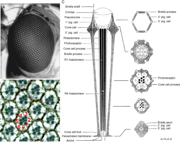
What type of cell it is?

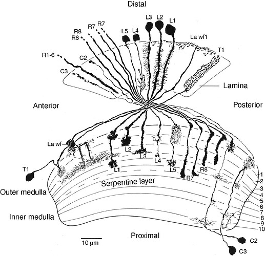
Going from illustrations in:


(Thank you @AzureJay ) and https://www.pnas.org/doi/10.1073/pnas.2025530118
and
(source: https://www.cambridge.org/core/books/abs/behavioral-genetics-of-the-fly-drosophila-melanogaster/anatomical-organization-of-the-compound-eyes-visual-system/8BD880CF0E0AA68EF0782E238C9682D5)
I’m making a hypothesis that the shorter the length of the axon the smaller the number (R1-8), so I’m thinking:

R1
https://ngl.flywire.ai/?json_url=https://globalv1.flywire-daf.com/nglstate/4576471385374720
https://ngl.flywire.ai/?json_url=https://globalv1.flywire-daf.com/nglstate/6366956009553920
https://ngl.flywire.ai/?json_url=https://globalv1.flywire-daf.com/nglstate/6335490299002880
https://ngl.flywire.ai/?json_url=https://globalv1.flywire-daf.com/nglstate/4774040048762880
https://ngl.flywire.ai/?json_url=https://globalv1.flywire-daf.com/nglstate/5683473268342784
https://ngl.flywire.ai/?json_url=https://globalv1.flywire-daf.com/nglstate/6042194339168256
https://ngl.flywire.ai/?json_url=https://globalv1.flywire-daf.com/nglstate/4718810787479552
https://ngl.flywire.ai/?json_url=https://globalv1.flywire-daf.com/nglstate/5698150840926208
https://ngl.flywire.ai/?json_url=https://globalv1.flywire-daf.com/nglstate/4932346092453888
https://ngl.flywire.ai/?json_url=https://globalv1.flywire-daf.com/nglstate/4627705001672704
https://ngl.flywire.ai/?json_url=https://globalv1.flywire-daf.com/nglstate/5353413789351936
https://ngl.flywire.ai/?json_url=https://globalv1.flywire-daf.com/nglstate/5707995577057280

R2
https://ngl.flywire.ai/?json_url=https://globalv1.flywire-daf.com/nglstate/5773884804562944
https://ngl.flywire.ai/?json_url=https://globalv1.flywire-daf.com/nglstate/4779279741091840

R3
https://ngl.flywire.ai/?json_url=https://globalv1.flywire-daf.com/nglstate/6603627397382144
https://ngl.flywire.ai/?json_url=https://globalv1.flywire-daf.com/nglstate/5658768952000512

R4
https://ngl.flywire.ai/?json_url=https://globalv1.flywire-daf.com/nglstate/4890495369084928

R5
https://ngl.flywire.ai/?json_url=https://globalv1.flywire-daf.com/nglstate/4959581126000640

R6
https://ngl.flywire.ai/?json_url=https://globalv1.flywire-daf.com/nglstate/5731199280480256

R8
https://ngl.flywire.ai/?json_url=https://globalv1.flywire-daf.com/nglstate/5973594072940544
https://ngl.flywire.ai/?json_url=https://globalv1.flywire-daf.com/nglstate/6332962576531456
https://ngl.flywire.ai/?json_url=https://globalv1.flywire-daf.com/nglstate/5407152252387328
https://ngl.flywire.ai/?json_url=https://globalv1.flywire-daf.com/nglstate/5982268296265728
https://ngl.flywire.ai/?json_url=https://globalv1.flywire-daf.com/nglstate/6540437120614400
https://ngl.flywire.ai/?json_url=https://globalv1.flywire-daf.com/nglstate/6481381748113408
https://ngl.flywire.ai/?json_url=https://globalv1.flywire-daf.com/nglstate/5681822717444096
https://ngl.flywire.ai/?json_url=https://globalv1.flywire-daf.com/nglstate/6536544026361856
https://ngl.flywire.ai/?json_url=https://globalv1.flywire-daf.com/nglstate/6749139379421184
https://ngl.flywire.ai/?json_url=https://globalv1.flywire-daf.com/nglstate/5844710694322176

R1 or 2?
https://ngl.flywire.ai/?json_url=https://globalv1.flywire-daf.com/nglstate/6217767770390528
https://ngl.flywire.ai/?json_url=https://globalv1.flywire-daf.com/nglstate/4643243253825536
https://ngl.flywire.ai/?json_url=https://globalv1.flywire-daf.com/nglstate/5618275832561664
https://ngl.flywire.ai/?json_url=https://globalv1.flywire-daf.com/nglstate/5769143160668160
https://ngl.flywire.ai/?json_url=https://globalv1.flywire-daf.com/nglstate/6750594727411712

R5 or 6?
https://ngl.flywire.ai/?json_url=https://globalv1.flywire-daf.com/nglstate/5624694820569088

R2?
https://ngl.flywire.ai/?json_url=https://globalv1.flywire-daf.com/nglstate/5898259407044608
https://ngl.flywire.ai/?json_url=https://globalv1.flywire-daf.com/nglstate/6085481032843264
https://ngl.flywire.ai/?json_url=https://globalv1.flywire-daf.com/nglstate/4653096177238016
https://ngl.flywire.ai/?json_url=https://globalv1.flywire-daf.com/nglstate/6561627683946496

R3?
https://ngl.flywire.ai/?json_url=https://globalv1.flywire-daf.com/nglstate/6744175739404288
https://ngl.flywire.ai/?json_url=https://globalv1.flywire-daf.com/nglstate/5356783619014656
https://ngl.flywire.ai/?json_url=https://globalv1.flywire-daf.com/nglstate/4959399260979200

R4?
https://ngl.flywire.ai/?json_url=https://globalv1.flywire-daf.com/nglstate/5226430531108864

R5?
https://ngl.flywire.ai/?json_url=https://globalv1.flywire-daf.com/nglstate/5139421338796032

R7?
https://ngl.flywire.ai/?json_url=https://globalv1.flywire-daf.com/nglstate/5572314850656256
https://ngl.flywire.ai/?json_url=https://globalv1.flywire-daf.com/nglstate/5418628572774400

R1-3s?
https://ngl.flywire.ai/?json_url=https://globalv1.flywire-daf.com/nglstate/4511989053259776

R6-8s?
https://ngl.flywire.ai/?json_url=https://globalv1.flywire-daf.com/nglstate/6544528479617024

Lines 25-71 in gsheet.
3 Likes

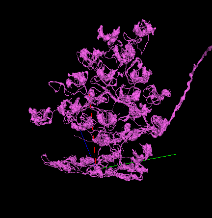
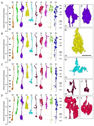
This is a Dm4 definitely - when they reach the edge of the lobe, the arborizations get squished and deformed, but still maintain the structure of having two main segments wrapping around a column.
An example on the other side of the lobe:
image
https://ngl.flywire.ai/?json_url=https://globalv1.flywire-daf.com/nglstate/6621195952717824

6 Likes

since the R8 is shorter than R7 in your picture i think that theory might be broken, could ofc be that they found R8 after naming the other based on length but would not take a chance on that.
From what i can see in the fishback paper R1-6 i short not going into medula while R7 and R8 go into medulla with R7 beeing the longest one.
I am not sure if R1-6 is the name of the cell or just that it is not possible to see the difference on the different short retinal axons looking at the optic lobe.

3 Likes

maybe, but then the theory could hold for r1-6, 8 and not 7? lol idk.

1 Like

I dont remember which thread it was but someone suggested maybe having a different/new lightbulb colour for a cell that is completed and has been given an identification, what if it’s not a new colour but a white (or other colour) check or exclamation point on top of the green light bulb? Altho it is very small and idk to what extend itd be visible.

2 Likes

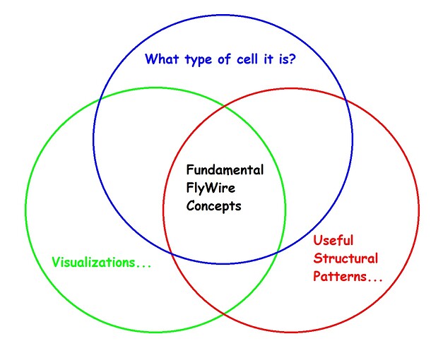
@Krzysztof_Kruk I have spent some time considering your suggestion, and I wanted to give you my take on the spontaneous and natural course that the discussion has followed, so far, using a familiar analogy,

"What type of cell it is?" and Nseraf’s identification gsheet - a particular, individual tree in the forest.

"Visual Cell Type Illustrations" and annkri’s examples gsheet - a visual catalog of tree types.

"Useful Structural Patterns…" - general patterns exhibited by the trees, and how they make up the forest as a whole.

I would propose that there is benefit in allowing the dicussion to follow its natural course, along the same lines that spontaneously developed, without planning or preconception. With that said, if we were to go back and write a book on the optic lobe, after the dataset has been completed, then we would most likely organize the book in precisely the way that you suggested, and in that sense you are absolutely right for suggesting it. It is perhaps worth noting, however, that the book is written “after the fact”, whereas our current discussions are taking place “prior to” and concurrently with “the fact”.

I am offering this merely as a way of conceptualizing things at present. There will, of course, be some overlap in the discussions from different topics, as illustrated in the figure below.

Cheers.

Cc: @AzureJay

5 Likes

@AzureJay My suggestion would be to hold off on uniquely identifying Tm/TmY cells for now (except for the really obvious ones, like Tm1, Tm2, etc.), until we have complete examples of each in annkri’s spreadsheet. We have not yet located and identified specific examples of each in our dataset. For now, as per @M_Sorek 's suggestion, we could just identify them as “Transmedullary” (i.e. without a number designation) when we are unsure…

…again, just a suggestion.

Cheers.

5 Likes

@Nseraf I’ve been wondering that myself…if there is some criteria for distinguishing the type R’s from one another (other than the obvious length difference between R(1-6) and R7 & 8). Before I go take a closer look, are you suggesting that you have observed a consistent length difference between R1 thru 6? If so, then you have proven your genius yet again sir, lol, as that would be exactly the kind of criteria we needed! :smiley:

2 Likes

I found identifing Tm and TmY quite easy for most types. I’m usually focusing only on the medulla part and the rest is just for confirming/disproving the finding. I always start with the number of arborizations in the medulla. It quickly eliminates about 3/4 of the candidates. Then take a look, where are the arborizations (in which layers) and how do they look. It works for about 90% of my identifications.

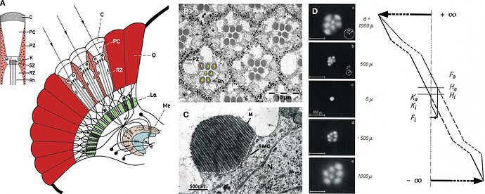
I was searching for some informations about the R cells, and found this paper:

According to the paper, the cells are oriented this way:

R1-R6 make this trapezoid shape, R7 is in the center and R8 is directly under R7 (under the whole cell).

From the same article:
" The R1–R6 cells (Figure 1 B) project their axons to the first optic lobe, the lamina (La, Figure 1 A green). The second group consists of two cells in the center of each ommatidium termed, R7 (marked 7 in Figure 1 B) and R8 (located below R7) each spanning only half of the retina in length."

I also found this image:

Here we can see, that the R8 cell is wholly below R7.

So, taking into account all that information above, my conclusions are:

  1. All the cells, that go to the medulla, are R8 cells.
  2. Fishbach is wrong about the R7.
  3. Without seeing the retina, we won’t be able to tell, which cell from the R1-R6 group is which.

Edit:
And there are these types of drawings:

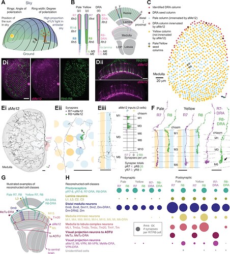
Source: Synaptic targets of photoreceptors specialized to detect color and skylight polarization in Drosophila | eLife

Which confuses the heck out of me :smiley:
It shows both, that R8 starts, when R7 ends, and that both reach the medulla. Ergo: more research needed :smiley:

Edit2:
Even more confusion from the last article:
" In the retina, inner photoreceptor R7 (distal) and R8 (proximal) rhodopsin expression differs across three functionally specialized subtypes pale (p ), yellow (y ), and DRA (d ). Rh3 and Rh4 opsins are both UV-sensitive, whereas Rh5 and Rh6 are more sensitive to blue and green wavelengths, respectively. R1-6 express Rh1 and are broadly sensitive to green and UV wavelengths. Only in the DRA, both R7 and R8 axons terminate in the same layer of the medulla neuropil (M6 ), which is the target layer of R7 cells outside the DRA, and non-DRA R8 cells terminate in layer M3."

4 Likes

I think we will probs end up breaking the R’s into 3 groups. General putative R1-6 batch, R7s which are the longest of them all, and R8s which are longer than 1-6 but shorter than 7s. 7s & 8s we will be able to be certain of/abt and 1-6s will always be putative not b/c we wont know/ be cecrtain they are R’s but b/c we wont be able to be certain if its a 1 or 2 or 3 etc. so probs:

Retinal R putative 1-6s

Vs

Retinal R7/8s

that’s my conclusion, lol.

4 Likes

We can’t be sure, if we have an R7 or an R8 sensitive to DRA. So we can only be sure about non-DRA R8 cells, and even that only, if we identify correctly the layer in medulla xD (look at my second edit).

4 Likes

unless length is not a way to differentiate between 7 & 8, we can differentiate between 7 being longer and 8 based on 8 being shorter than 7, no?

3 Likes

nope, there are 3 sub-types of R7 and R8. The types are sensitive for pale, yellow and DRA (whatever it is), so we can have p-R7, y-R7, d-R7, p-R8, y-R8 and d-R8.
All R7 and d-R8 go to the sixth layer. Only p-R8 and y-R8 go to the third layer of medulla.
So we can be sure only about p-R8 and y-R8.

4 Likes

Ah…how lovely…

3 Likes

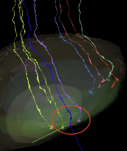
So how many of these would you say are 8s or 7s?
https://ngl.flywire.ai/?json_url=https://globalv1.flywire-daf.com/nglstate/4592115963133952

3 Likes

I’d say, at least 3 (the shortest ones) are R8.
The rest are either R7 or d-R8.

3 Likes

ok, so 2 batches, putatives R1-7s and R8s lol

2 Likes

Rather three:
Putative R1-R6 (all that don’t go into medulla)
For sure R8 (all that go to the third layer of medulla)
Putative R7 or R8 (all that go to the sixth layer of medulla)

3 Likes

i would say the three circled might me Dra, but do not really think they are, the rest are either pale or yellow so you can see it on the length. According to the last illustration i kk´s post Dra clusters are only a single line on the edge of medulla. also according to that picture it looks like the Dra clusters might have a bit more volum at the end compared to the other types.

I have not worked a lot with R cells, but could that macro structure with R1-R6 going together in a bundle still be visible in our dataset? if not only way to identify them wouldprobably be where they have the synapses.

4 Likes