I’ve tried to find a bundle. There were some bundles of 7 R cells, but the R7/R8 wasn’t in the middle and the whole thing didn’t have a trapezoid shape. So not sure, if it’s possible to differentiate them that way. However, I’ve only checked 2 such bundles.
The different endings and the synaptic points are both good ideas to check.
@Krzysztof_Kruk @Nseraf @annkri
I have another suggestion, for a possible criteria to distinguish the R’s from one another…
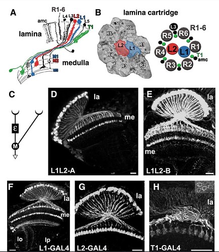
In my experience, the L3 cell is almost always located between two of the incoming retinal axons. If, for example, it turns out that it is always located between the same two retinal axons, and if the axons are always consistently numbered in a clockwise or counterclockwise fashion, then we could in principle determine some or all of the axon numbers based on that…
…note that there are two very big “ifs”, in the suggestion above. ![]()
@Krzysztof_Kruk In your research, have you come across any mention of the axon orientation, relative to the L3 cell of the cartridge?
No. I only found out that at the larval state (or maybe it was a pupa), in the center there are L1 and L2 and the R1-R6 are around them. That changes, when the fly grows up.
@TR77 According to your research (which I agree with), the cell in the gsheet marked as Mi8 looks like a Mi9. Could you confirm? And if so, past the correct link.
@Krzysztof_Kruk Sure…let me take a look. Also, regarding the R orientations relative to the L3 cell…AJ has two illustrations in “Visual Cell Type Illustrations” that both show the L3 cell located between R5 and R6, from two different sources (possibly). The illustrations I’m talking about are up near the top/start of the thread…give me a minute and I’ll paste the photos here, for reference…and I’ll go take a look at that Mi8/Mi9 issue…
@Krzysztof_Kruk Yep, lol, that’s definitely Mi9…been staring at those things for days now, lol. Good call, and nice catch, KK. I’ll go fix it…
Thanks ![]()
Yes, I’ve also seen it many times. Only now, when I had trouble identifing my cell (Mi8 vs Mi9), I’ve spotted that they look very similar. Then found your entry about those and it raised my concerns ![]()
As for the two pictures, they both are from articles about developing brains (the thing I said before - L1 and L2 in the middle with ring of R1-R6 around them, no R7 or R8). I wonder, if much changes during the development state in regards to the other cells and where L1 and L2 go.
But we could definitely search for structures containing L3 and some Rs and then try to find the rest of the Rs. Maybe it’s always the same. After all, L3 is a little “grabby” with all those synapses, so when it catches something, it probably holds it for life ![]()
@Krzysztof_Kruk Nice! …that was a team effort, and a win as a team, on the Mi8, lol. ![]()
And yeah, the L3 thing seems worth looking into… I honestly haven’t spent any time thinking about it until now, when I saw you all discussing the type R’s here, so… I’ll look into it a little too, and see what I come up with.
Cheers.
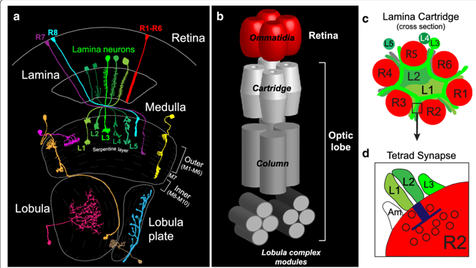
@Krzysztof_Kruk Oh hey…I can tell you for sure that the ring of R’s around a laminar cartridge is preserved in our dataset (as confirmed in the link @annkri just posted above). That is, after all, what the laminar cartridges are doing…synapsing with the retinal axons…
Indeed, I was just writing, that the L1 and L2 cells are still there, probably just lower in an adult brain, which is greatly visible in @annkri’s link. So now we have to find, where are R7 and R8 from that cluster and if they are somewhere else, then we can be quite sure, the ring is made from R1-R6. The order probably doesn’t change from larva to imago, so we would only have to find a way to tell, where the ring starts and in which direction it’s going. Probably your finding about L3 could help greatly.
I love, when everything falls in into place ![]()
warning: 282 cells ![]()
defo some fully traced clusters in there tho ![]()
That’s become my plan of attack moving forward, but I also thought I’d post some of remaining ones I had left in my ID log in case anyone’s got a clue. I am leaning into M’s suggestion that we focus on obvious IDs only as we’ll come back later through the data set to really dig down into identifying once we’ve mapped everything out.
As we can see, a lot of Identification is going to rely on our synapsing partners, reviewing the entire cartridge for relativity to each other, etc.
This is a cool idea! I like the idea of a community naming discussion. Something like this will definitely happen once the brain is complete but that could be a while. @annkri is right that it is possible to query cells that haven’t been IDd, and one could sort by user or groups of users or also by primary input and output neuropil. In the mean time, we could try to set up some community chats with scientists or even an optic lobe town hall if that would be of interest!
You can resubmit the correct cell identification. Both IDs will show up on the neuron. We don’t have a good way to clean the data yet but at least if you submit a second, accurate ID, it will remain attached to that cell.
How cool about a new cell subtype!!
I’ll circle back if the scientists have any thoughts on identification labels.

