As a prelude to a guide, the link below contains several completed and identified L1 cells, enough to give a basic impression of the arrangement of the lamina (each cell being the center of a laminar cartridge). Just imagine several hundred more, filling in the gaps, and you will have the right picture in mind.
The first image below, shows the “clumps” of L1’s and L2’s that I mentioned in a previous post. In the EM images, they appear as lighter areas, surrounded by darker areas, with each lighter area being the center of a cartridge. In the second image below, the cartridges present something of a “honeycomb” appearance in the EM, in some spots, which makes them really easy to spot…again, each of the honeycomb units being an individual cartridge.
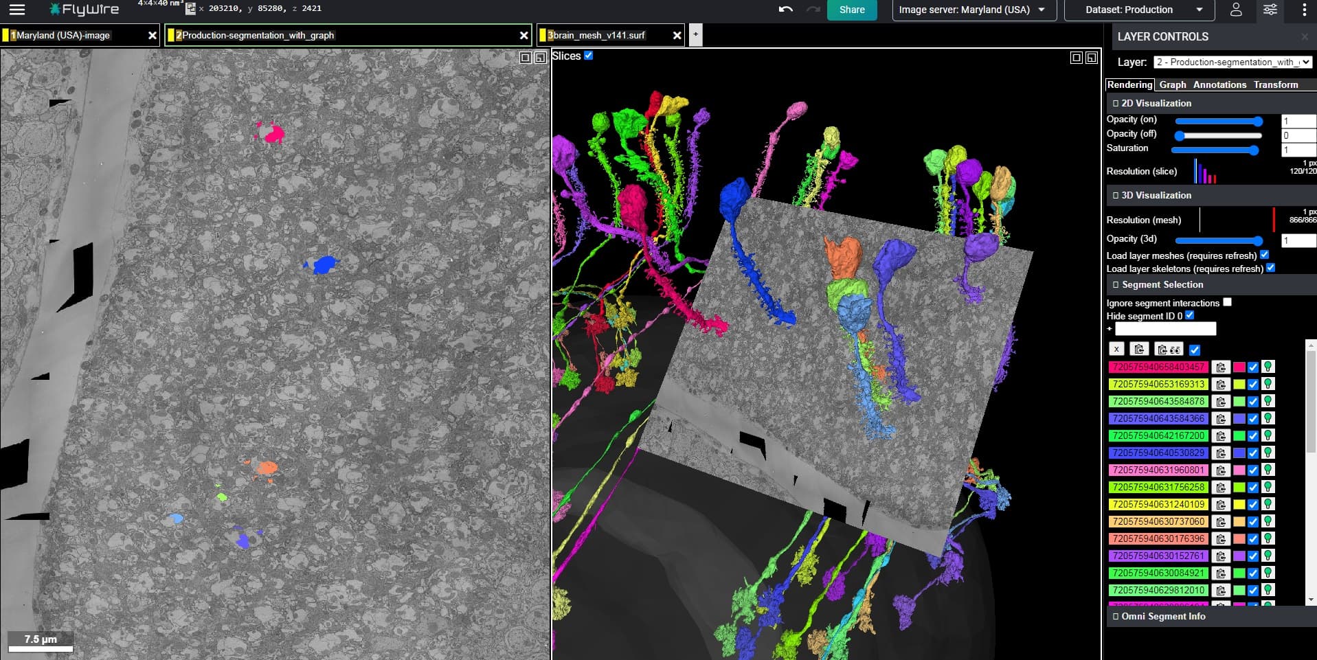
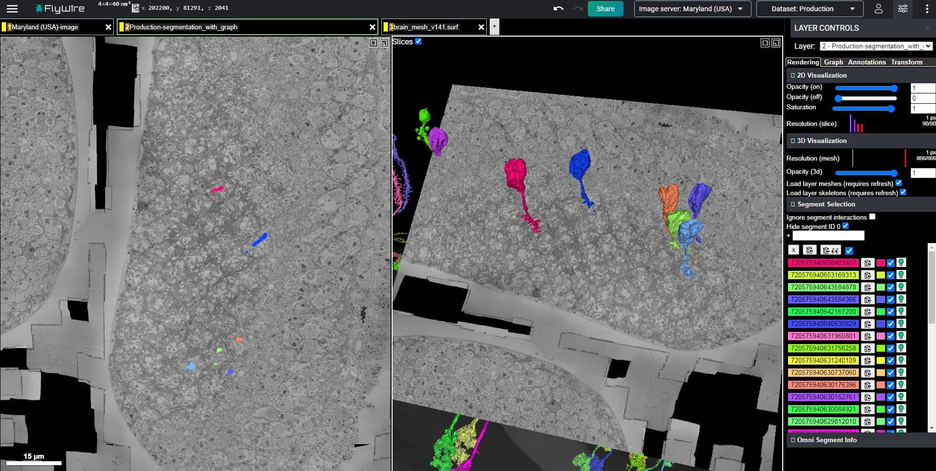
Finally, this link from the FlyWire blog shows all type L cells of a single cartridge together (i.e. L1, L2, L3, L4, and L5). You can add in the T1 cell with Id 720575940619753307 which forms something like a “basket” or “cage” around the cartridge. There are, of course, other cells associated with each cartridge, but those six cells represent the basic structural unit of the cartridges, and should give you a good idea of what you are looking for.
This is incredibly helpful, thank you! I look forward to the full guide but I think this is a very useable start to get pecking at the tricky cells that have plagued us.
Incredible work! If y’all need some more L cells to look at here’s a link of a few of them: https://ngl.flywire.ai/?json_url=https://globalv1.flywire-daf.com/nglstate/6701824207749120
1-2 of them are from q/a sheet w/o cells (my cells im looking for their CBs) but other than that they’re all complete. There’s also some retinal axons in there but they synapse and/or ‘Cartridge’ with L cells so I left them in.
a good idea making a guide, in my option it would be really useful to add in pictures that could be used as a printed out version + the 3d links if needed. Since i am tracing on one screen only looking at it on the computer is less useful than having it on paper. And not too many cells at the time since that is making the screen more messy.
The type L cells form their own well defined and exclusive CB layer, so…I know it goes without saying but, you can always just start randomly clicking on nearby somas, and you will light up L cells all over the place, lol.
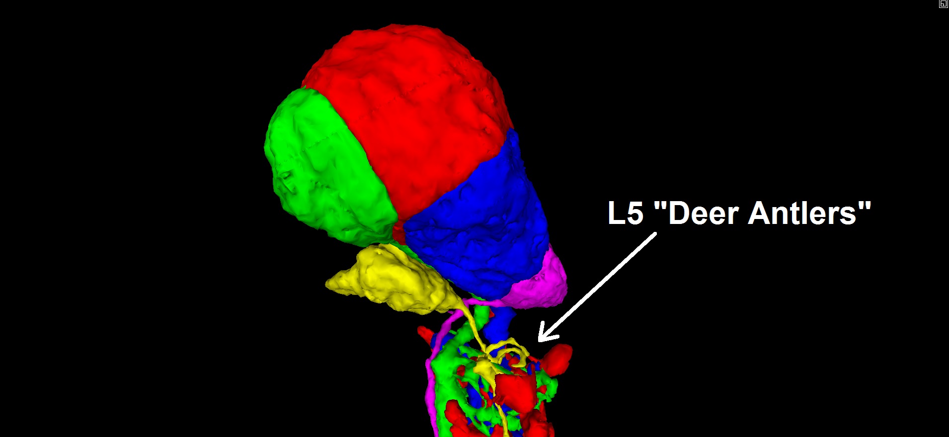
The L1, L2, and L3 somas (blue, red, and green below) tend to mush together into a clump, while the L4 and L5 somas (magenta and yellow below) tend to be smaller somas, located just beneath the clump of the first three. Also, the L5 cell seems to always have a small “deer antler” arbor, immediately downstream of the soma, which can help to identify “pinched” L5 segments with the soma attached (have seen a few instances in the Q&A log).
Here’s a ‘full’ circuit (I think), FlyWire and annotated, L’s, T’s, retinal axons, etc. at least for the ‘top’ part, the ‘lower’ part’s a mess of many more types, lol.
Now im wondering if the LAWF’s are taking info from the L’s and T’s and sending it elsewhere and T’s and L’s take info from retinal axons or the pathway is diff.?
the ‘other’ are glia, and other stuff not -attached- to something else larger, standalone branches etc.
Maybe we (the players) should create a sheet in the spreadsheet with all types of neurons listed (glia can be there too ) and try to find at least one example for each type. It would be both a challenge and a helpful tool for newer players and those less knowledgeable in the field.
@Nseraf Yeah, clearly a MASSIVE parallel processing layer (i.e. Photo-receptor > Retina > Laminar Cartridge > Medulla > … > Central Brain). It reminds me of things I have seen in industry.
@Krzysztof_Kruk I was thinking something along the same lines, as well. Rather than creating a guide specific to laminar cells, I would like to ultimately create a general guide to inferring microstructure from macrostructure (when the need arises, due to problems encountered in the EM data). I’m picturing a collaborative wiki-style document, where we describe useful macrostructural patterns, as we encounter them, and characterize the associated cell types (including useful recognition heuristics, such as the L5 “Deer Antlers” that I pointed out above).
At the moment, I would like to extend this concept of inferring microstructure from macrostructure, based on structural regularity, to the Medulla, and then on to the Lobula areas. If anyone has any observations that would be helpful, feedback would be most welcome.
When I was browsing the identified cells, I’ve noticed, that some of them are missing some structures.
Link: Sign in - Google Accounts
You can type “missing”, “lacking” or some other helpful keywords to find those cells.
I’m wondering, if we could find the missing parts. In some cases, it would be, of course, impossible, like in cases, where something is missing because of the dataset edge or missing segmentation, but some entries saying “missing branches” sound, like something could be done with it.
Keep in mind, some of them are in older segmentation, so you’ll have to click the red clock icon on the “Production segmentation” tab to update it.
Not sure, if it would help and if we should update the identification descrption after we find something. So it’s a post just saying, that such things exist.
EDIT: we could use that page to find cells of types missing in the spreadsheet
I’m not sure we can edit those cells, I tried to add an extension to the last one on the link and it tells me “merge does not work with a segmentation at an older state” and the same happened when I tried to remove a merger, “split does not work with a segmentation of an older state”.
But having said that, even if we can’t edit them, maybe if one of us does find a valid connection between one of those cells and the cells in the q/a sheet an admin can merge them together and complete the cell(s).
into the source field, then click inside the Name field and then save.
After that you can switch to the new tab and add fragments from 1 to 74 to see various parts of the brain in different colors.
You can change the 3D opacity of this layer and combine it with some cells to get some nice pictures.
Fragments, you may be more interested in, are: 2, 6, 14, 29, 35, 43, 51, 56 (parts of the optic lobes).
@Krzysztof_Kruk Are you by any chance familiar with the syntax for querying that database (identified cells)? Any tips for us? It looked to me like there is at least SOME inconsistency in the naming of identified cells, in that database (haven’t actually verified that yet, though).
Oh and, awesome tip about the neuropil meshes…thanks!