Useful Structural Patterns in the Optic Lobe (inferring microstructure from macrostructure)

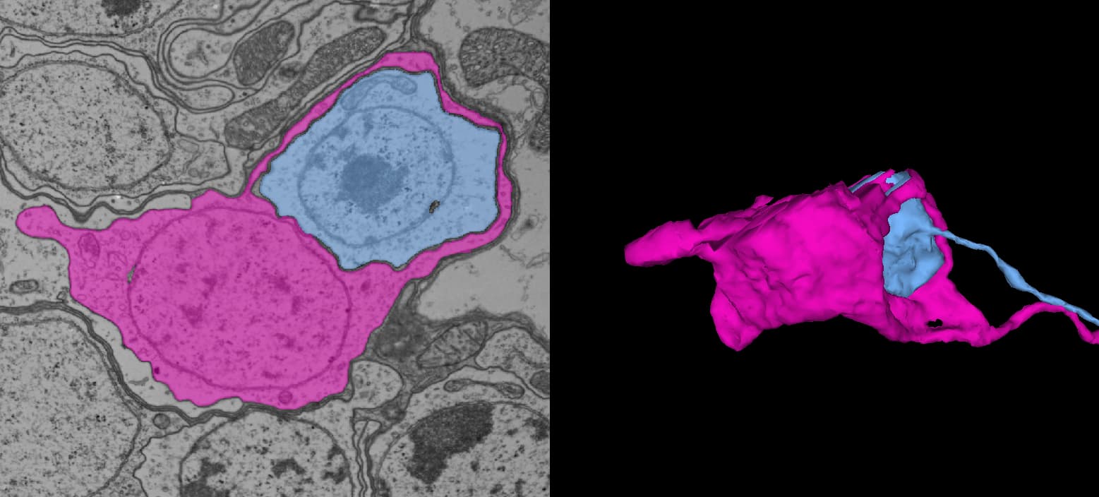
I decided to go ahead and create a thread solely devoted to this topic, as it has proven to be a very useful and practical approach to dealing with some of the issues encountered in the EM imaging data. I do realize that most of what is presented below will already be familiar to most of you. But, I felt that it would be worthwhile to document our observations here, in a separate topic, as a reference for ourselves, and as a primer for the benefit of newcomers.

Inferring Microstructure from Macrostructure

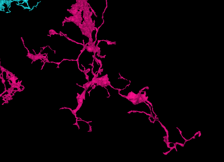
So, what do we mean by “inferring microstructure from macrostructure”? Well, suppose that you own a small orchard, of hypothetical trees that always grow in the same way, and always have three branches. The trees in your orchard are arranged in perfect geometric rows, with each tree being a nearly identical copy of every other tree. Now suppose that you go for a walk in your orchard, one day, and you come across a peculiar tree with only two branches, and find a dead branch laying on the ground beside it. It would be perfectly reasonable to “infer” that the dead branch on the ground is, in fact, the missing third branch of the peculiar tree. This would be an example of “inferring microstructure from macrostructure”.

Columnar Structures and Row Fields

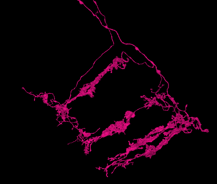
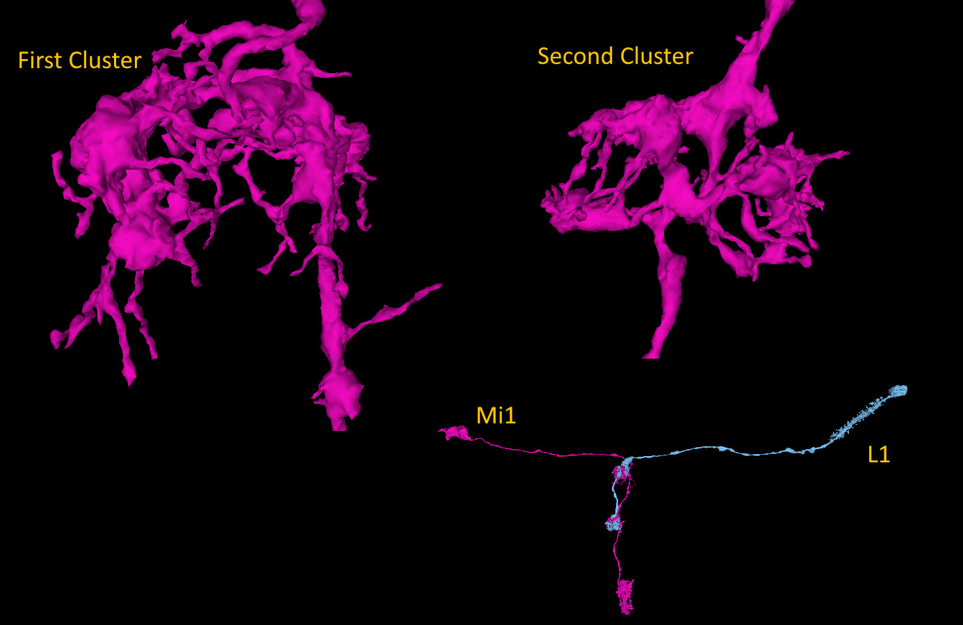
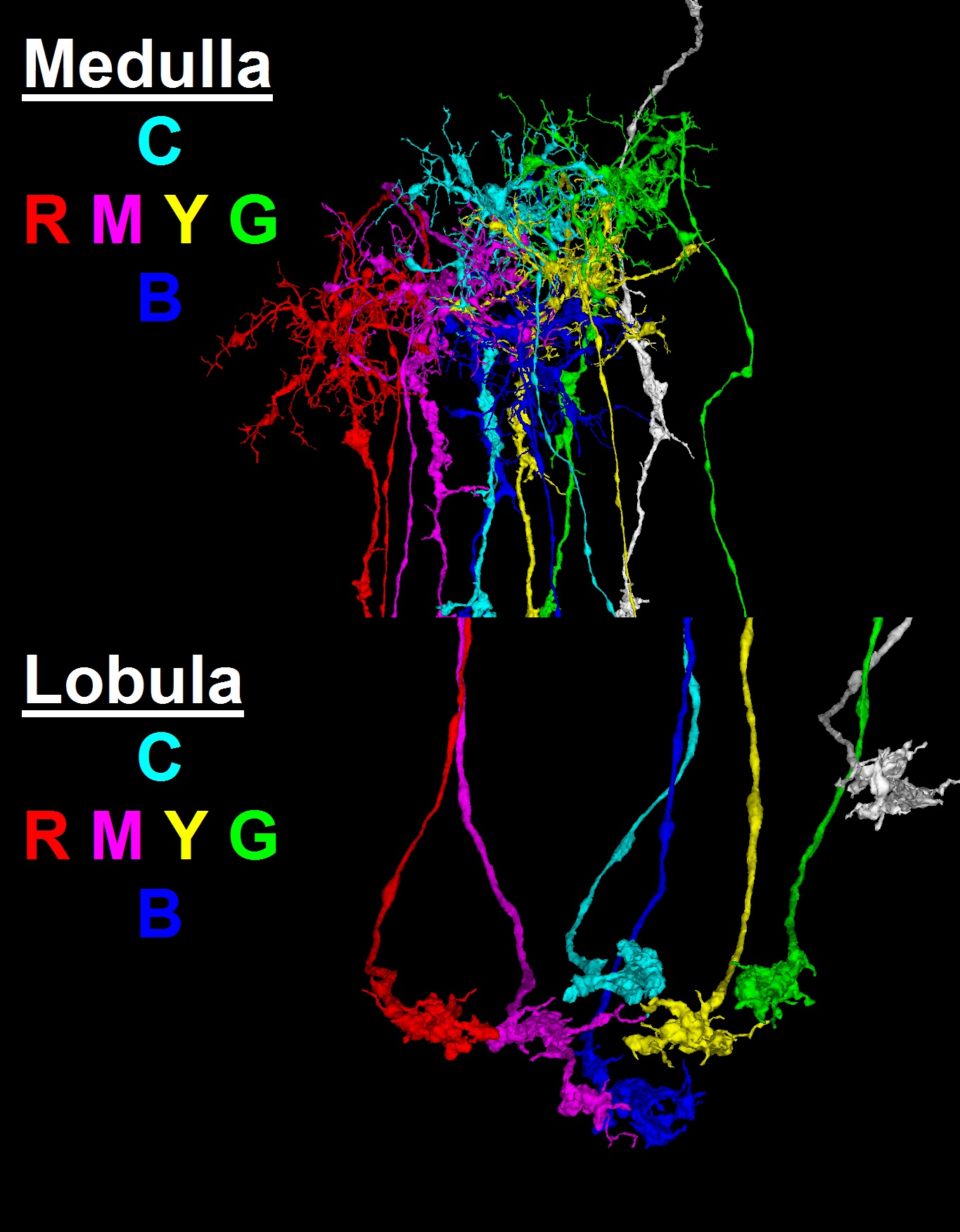
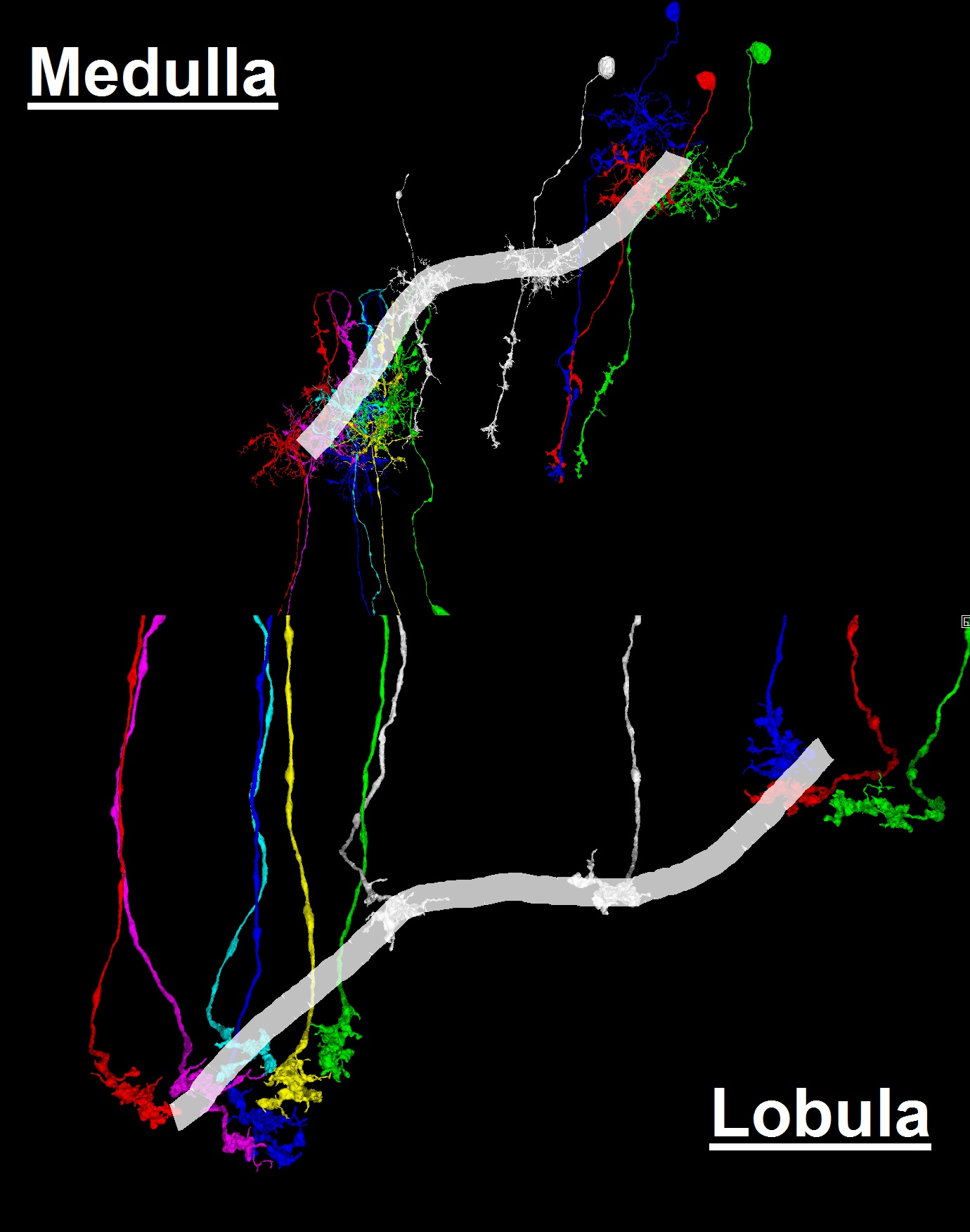
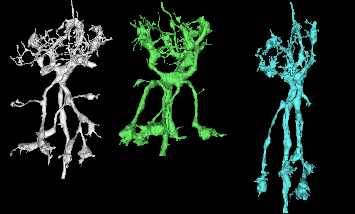
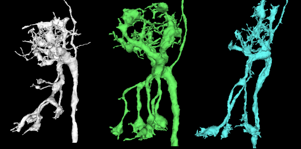
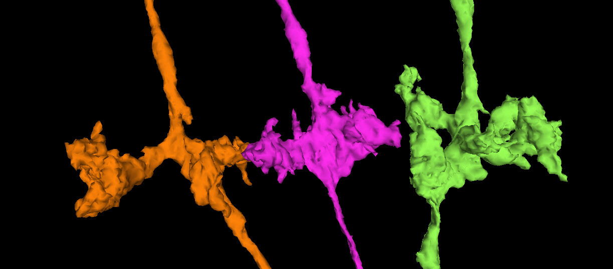
The link pictured below (as a clickable YouTube video thumbnail) contains a collection of neurons selected from the “Zone 2” set (in the Flywire blog), and several additionally selected neurons. This selected group of neurons is presented here as being representative of the general overall structure of the optic lobe. As can be seen, the structure of the optic lobe can be characterized as the combination of an array of regularly spaced and repeated columnar structures, with a series of several successive layers of perpendicular row fields, intersecting the columnar array. A familiar example of the columnar structures of the optic lobe, would be the L cells of the ubiquitous “laminar cartridges” (found repeated throughout the lamina), which represent the “tops” of the columnar structures of the optic lobe. A familiar example of the well defined row fields of the optic lobe, would be the Dm cells (found repeated throughout the medulla), which have been featured in the “Gallery of Amusements” topic, for their aesthetically pleasing appearance, which sometimes resembles a field of flowers.

Applying the above, as an approach…

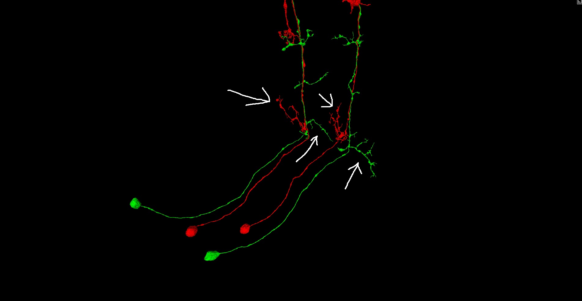
Recognizing the overall general structure of the optic lobe, as well as the component substructures, can be helpful in anticipating where and what to look for, when searching for the extensions of a particular cell, or group of cells. For example, if your attention is called to a particular L cell in the lamina, then without even looking, you can predict it’s soma location, arbor locations, and overall morphology, as well as anticipate that it neighbors and interfaces with several other L cells in a predictable way, along with several R’s, T’s, C’s, etc. This consistency and predictability of structure can be of tremendous value, when problems in the EM imaging data are encountered.

In closing…

Feel free to post here as much as you like, as the search tool can always be used to locate posts relevant to a particular cell type or structure. A “useful structural pattern” could be anything from noticing consistency in the shape of a particular cell type’s arbors, to noticing that a small group of cell types almost always occur together and in the same arrangement, and even noticing large scale structural patterns related to the overall structure of the optic lobe.

Cheers, all.

https://ngl.flywire.ai/?json_url=https://globalv1.flywire-daf.com/nglstate/5451622914195456

5 Likes

I’ll go ahead and start this off with a collection of links to my previous posts, from other topics, relevant to the “laminar cartridge” structures of the lamina.

Keywords: lamina, cartridge, pinched, black spill, L, L1, L2, L3, L4, L5

"Pinched" Neurites, and the Infamous Row 4 Cell - a post about the reconstruction of type L cells whose backbones appear to have been “pinched” apart.

A post about recognizing and locating L cells in the lamina.

Another post about recognizing and locating L cells in the lamina, with graphic illustrations.

A post about the somas and CB layer of L cells.

Cheers, all.

:heavy_minus_sign: :heavy_minus_sign: :heavy_minus_sign: :heavy_minus_sign: :heavy_minus_sign: :heavy_minus_sign: :heavy_minus_sign: :heavy_minus_sign: :heavy_minus_sign: :heavy_minus_sign: :heavy_minus_sign: :heavy_minus_sign: :heavy_minus_sign: :heavy_minus_sign: :heavy_minus_sign: :heavy_minus_sign: :heavy_minus_sign: :heavy_minus_sign: :heavy_minus_sign: :heavy_minus_sign: :heavy_minus_sign: :heavy_minus_sign: :heavy_minus_sign: :heavy_minus_sign: :heavy_minus_sign: :heavy_minus_sign: :heavy_minus_sign: :heavy_minus_sign:

Please take a moment to respond to the following poll. Your feedback is valued and appreciated.

Which of the following statements best describes your reaction to this forum topic:
  • This topic is wonderful, and I find the information very helpful and useful!
  • I’m a mean person, and I still wear baby diapers.

0 voters

5 Likes

That’s…a lot of well structured info! Great work!

An observation i’ve had is that somas from same types of neurons are usually (if not always) in the same soma layers, so if one has a backbone from a type of cell s/he/they recognize (ie a T or an L or a Dm etc) and they can find the soma/CB (black spill area etc) then scrolling up to the CB layer and clicking around discarding somas that have backbones or finding backbones that are not the ones you want may eventually yield you the soma you need. At least if you know that type’s soma layer in the dataset.

4 Likes

Distal Medulla 9 (DM9) *

The DM9 cell features two main parallel branches. The ‘lower’ branch is a small arbor cluster of boutons and dendrites.

The upper branch runs parallel to the lower branch. It has a wider spread, and features one or more drooping clusters of boutons (they look like grape bundles; one might think of them as rachis), one of which will intersect with the lower branch.

The entire structure is fairly linear, with the upper branch continuing straight out from the soma.

Known examples (if you have more, please let me know and I will add them and update the link): FlyWire

*To the best of our knowledge this is the believed type of cell.

5 Likes

think I found another one: 720575940627420164

3 Likes

@AzureJay Awesome, AJ! I really like the idea of keeping an updated link with all known examples of a particular cell type. If we had that for every cell type, then we would have like a virtual library of information! Perhaps we could include links like this in @annkri 's cell structure example spreadsheet?

1 Like

I was thinking about it when starting the sheet, but my conclusion was that it would be way too much work. And that you also run in danger for adding the wrong label if everyone could add cells without really knowing what they are doing (i am only adding cells labeled by scientists where i can find that)

But if anyone would like to have a link like that i am thinking we could add a new column at the end with a link where we can add additional cells?

5 Likes

@annkri Yeah, that sounds great! :smiley: And good idea, separating the documented cell type examples from the community identified examples. I’m all for adding a new column, where we can keep an updated link of several community examples. Also, it wouldn’t have to be ALL known examples, as that could become rather tedious. I’m thinking maybe a dozen or so examples, from different locations in the dataset…enough to give an impression of the structural patterns, for a given cell type (i.e. soma locations, arbor locations, arbor shapes, paths followed, and overall morphology in general).

Great idea! @AzureJay and @annkri :100: :bangbang:

4 Likes

Agreed on the idea of “Scientist Verified” versus “Community Link” on the spreadsheet, and yes, let’s keep it to maybe a dozen examples at most particularly for those who have a little lower internet speeds.

If my DM9 post was useful I will try to make more like it in the future for other cell types. I know many of us were puzzling over it in chat the other day so it seemed like a good starting point.

3 Likes

@AzureJay Yes, absolutely…more posts like your Dm9 post would be VERY welcome!

Cheers. :+1: :100:

1 Like

i added a link too the post about both Dm9 and L cells (first in discussion) in the sheet so it is easier to find when needed

3 Likes

@annkri Outstanding! Thank you! :smiley:

Okay, so…check out how awesome this is!

In the L1 cell entry, in @annkri 's “example of different cell structures” spreadsheet, you can now view community selected preview images (columns C thru E), view an officially documented example of the cell in our dataset (column F), read a brief community description of the cell (column G), view a link containing several community identified examples in our dataset (column H), and even click a link to a discussion about the cell in the community discussion board (column I)!

My goal now is to complete an entry like this for every cell type in the optic lobe…b/c that would just be fantastic, lol.

Cheers, all.

6 Likes

Medulla Intrinsic 1 (Mi1)

The Mi1 cell features a single branch with three total clusters - two dendritic, and one boutonic. The soma begins outside the medulla, and the branch takes a hard angle turn (approximately 90 degrees) into the medulla.

The soma has a ‘blanket’ layer which wraps around another cell’s soma (a TM type):

The first two “dendritic” clusters are roughly spherical. They wrap around and interface with an L1 cell.

The final “boutonic” cluster features several (3-4 seems to be common) bleb-type bundles that go upward along the axon:

Known examples: FlyWire

5 Likes

@AzureJay Outstanding work, as always, AJ! :star_struck:

3 Likes

Avesome post AJ
After working on the sheet, i see that the part i have had too little focus on is where in the brain you can find the cell. Partly because i did not see the importance of this at the beginning and have just continued using the same pattern for the pictures. And partly because it is very difficult to get good pictures showing this when it have to be visible on small pictures.
With a forum post i am thinking this would be a good place too show this,
maybe a short video zooming in/out and around the brain mesh might be a good way? maybe even use the coloured brain parts that amy have showed us, if that would show it better.

5 Likes

I’d just like to add, that the type of the structure seen on the last picture is sometimes called bleb-type arborization.

3 Likes

Miscellaneous useful structural pattern…

Noting here, a pattern that I have been seeing, in the arborization of Centrifugal C2 (green) & C3 (red) cells, at the location of the 90 degree bend. This pattern can be useful in identifying and reassembling fragmented segments of these cells. In other words, when you locate a soma with a piece of the backbone attached, you can often quickly determine which cell’s soma you have located, based on the pattern of arborization at the 90 degree bend.

https://ngl.flywire.ai/?json_url=https://globalv1.flywire-daf.com/nglstate/6193692196995072

4 Likes

Miscellaneous useful structural pattern…

Noting here (or rather confirming here), that the relative arrangement of Transmedullary Tm/TmY cells in the medulla, is preserved in the lobula. Specifically, in the diagrams below, the arrangement of the dendritic arbors of Tm4 cells, in the medulla, are shown in comparison with the location of their axonal arbors, in the lobula. As can be seen, the relative arrangement of the cells is preserved both locally, and over large areas as well. I have not verified this pattern for every single one of the many types of Tm/TmY cells yet, but have observed it in several already, and felt it was worth pointing out and confirming here.

This can be useful in locating and reconnecting separated pieces of these cells, in two different neuropils. Basically, if you know the location of the cell in one neuropil, then you already know the location of the rest of the cell, in the other neuropil.

As an aside, it is perhaps not surprising, in some ways, that the relative arrangement of the cells would be preserved in this way. The Tm/TmY cells, after all, are presumably transferring the entire visual field from the medulla, down to the lobula. So, it is perhaps of little wonder, that their relative “pixel locations” (if you will) are preserved in the transfer.

https://ngl.flywire.ai/?json_url=https://globalv1.flywire-daf.com/nglstate/5450687148195840

5 Likes

Transmedullary 2 (Tm2)

The Tm2 cell has a straightforward branch pattern with three total clusters - a dendritic arbor, and two small bleb-like clusters. It starts above the medulla near the edge of the EM dataset, passing through into the lobula along the corridor between the lobular plate and lobula.


The first cluster is an arbor found in the medulla and has a unique “X” type structure, with the top of the X having small dendritic twigs and the bottom having larger bouton extensions. This is the most recognizable part of the cell.

The second cluster, in the lower medulla, features only a couple bleb-like extensions.

The final cluster, found in the lobula, has only one bleb-like structure.

Example cells (as curated by @TR77): FlyWire

6 Likes