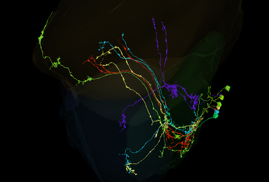
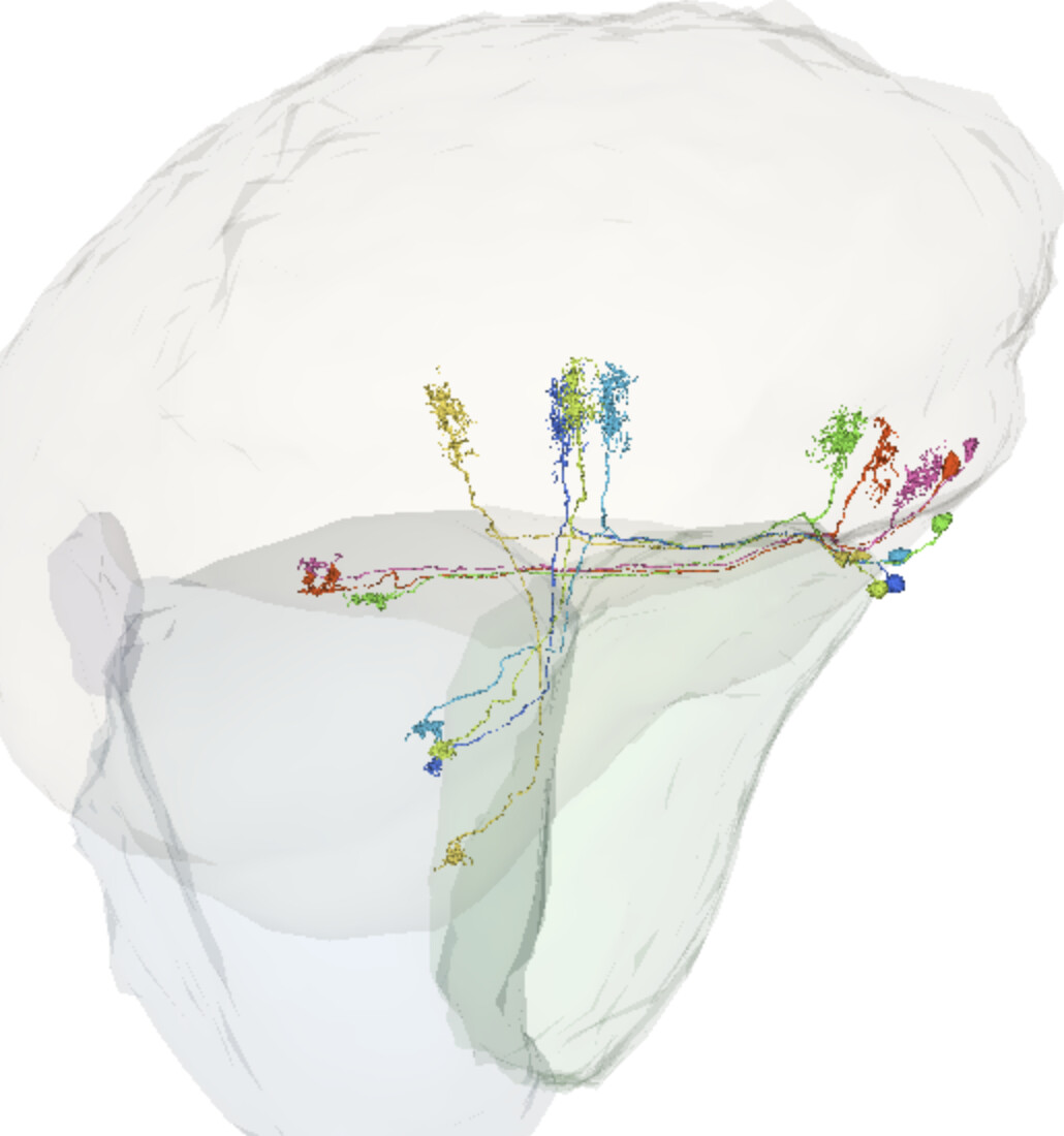
I have 8 cells here - FlyWire - from the outer lobula plate rind that I was not able to complete or identify in my pass (as described here). I would love your assistance!
I think a few of these are probably a Y type, but there’s confusing things all around here. One is probably centrifugal.
I’d say, 4 of them are Y.
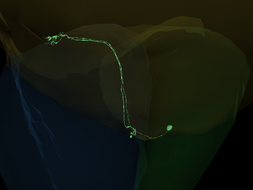
The short green one (065) might be an LPi.
The longest could be a merger (didn’t found any merging spot, though). The outer part has a missing extension, that looks like a C cell (I didn’t trace it to the end).
The one ending with 326 is unfinished. It’s continuation goes into a merger, but I didn’t check, what part of that merger is correct and if it continues beyond that, so it’s hard to say, for now, what type it is.
I don’t know, what to think about 218, but it also looks, like its unfinished.
Thanks for the feedback! I’ve done some updates and narrowed things down a bit:
Marked the four y cells as Putative Y and sorted them out. I had forgotten about Y11 and Y12 types which match them a lot more.
Edited two cells, one of which I’ve recompleted as Lpi type (720575940622000372, removed from link below). The other (-629 in the link) has been fleshed out a little more but I still feel unsure about it being complete, although it is probably Lpi too.
(-326) definitely feels like a merger, and I’ve added the opposite trace-back soma. I think it’s somewhere along the ‘break’ in the 3D, but I can’t put my finger on it still.
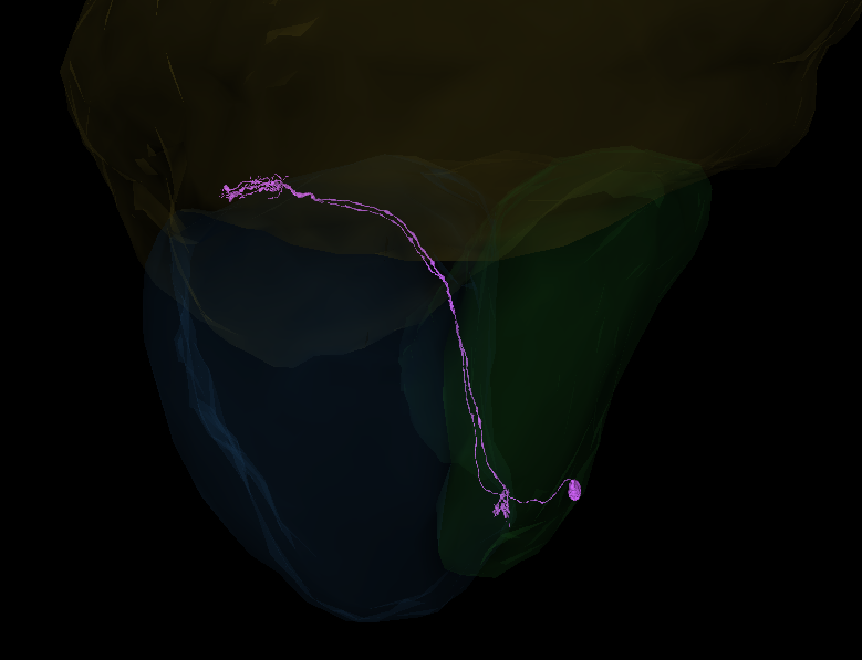
(-740), the extra long one, I haven’t been able to find anything more to point to a merger or an extension. The medulla end definitely reminds me of C-types but the rest of the structure doesn’t add up to anything. My best guess is that this is an LLPC that is merged with a branch that heads up to the medulla, and is missing its branch to the central brain.
Added the correct extension to -326. It’ll be a T cell after merging,
I think, the -629 looks complete enough,
Added more segments to -740. Now the top part definitely is a C cell. For now, I couldn’t find a merger either. I’m going to go to sleep now, so I’ll look at it tomorrow.
I’ve checked (more or less) the whole -740 and didn’t find any mergers. I’m starting to think, maybe there isn’t a merger. Maybe it is a C cell, but malformed. Maybe it grew in the wrong place at the wrong time and was dragged away from its normal position. Not sure, if it’s possible though
Thanks so much for the assistance and feedback, KK!
I’ve eliminated the T cell and Lpi now, which leaves us just with this stranger. I’ve merged together your additions, and made a few clean ups/additions myself where I could find them. I also combed through the 2D again, and I’ve marked two points that might be mergers. Though I will say even if they are mergers, we’re still drawing a blank on what this cell is!
I’ve tried looking at friend cells in different places, but not finding any clues. This bundle is mostly T5s, a few T4s, and one Tlp. I’m most inclined to think it’s an Lpi merged with something else, assuming it isn’t, as you suggested, a poorly grown ‘mistake’ neuron.
You’re welcome
Unfortunately, I can’t find any mergers either.
Maybe it’s a new kind od cell, some sort of probe connecting lamina with lobula and lobula plate. There aren’t any other cells directly connecting these neuropils, so maybe there’s a C cell repurposed just for this.Who knows. After all, we’re doing researching, so might find unknown unknowns
Quick guide, how to differentiate between T cells:
T1: Lamina → Retina T2: Medulla → Lobula (goes through the whole Medulla) T3: Medulla → Lobula (arborizes in the proximal part of the Medulla only) T4: Medula → Lobula Plate T5: Lobula → Lobula Plate ChaTnew1: Medulla → Lobula and Lobula Plate
The last one is also known as Tnew1 in some pictures.
Categorizing T cells definitely needs to have the optic lobe’s neuropils turned on, because they sometimes have their arborizations very close to the borders of the neuropils.
do you have a picture of the ChaTnew1 have not seen this before?
i am thinking on the differences more like this
T1= basket
T2= long arbour
T3= short arbour
T4 = turn 180 degree, goes into medulla
T5= turn 180 degree, goes into lobula
I was thinking about it. My T3 have its proximal end in the lobula neuropil, but I’m starting to think, that the 3D models of the neuropils aren’t exactly matching the 2D slides.
Yesterday I was sure, that they are correct, but the more time passes, the more I think, I got it wrong and the more I agree, that T3 should look only like the ones you posted.
At least, I didn’t post my classifications yet.
Agree about the neuropils not completly matching, making it even more difficult with the T4 and T5, from what i have observed i think there are many T4 cells that are just under the medulla neurophil, because of this i tend to classify all of the ones i am unsure about as T4
I wish, I could make the models more accurate and even add all the layers. Unfortunately, I don’t know how to.
Thanks, for straightening my knowledge about the T3s
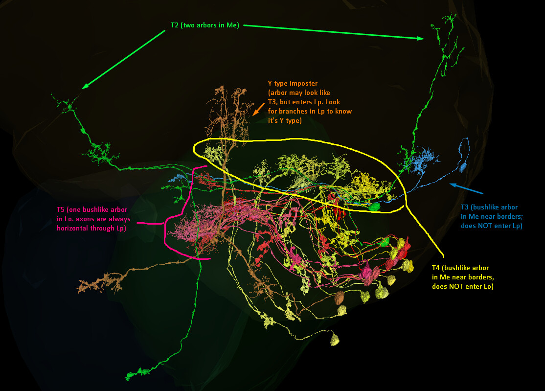
T2 (Green): always has TWO arbors in the medulla. Sometimes the first arbor will look similar to a T3 arbor, sometimes (like these examples) they will be more loose.
T3 (Blue): always has arbor in the medulla near the neuropil borders. Usually it is bush like but it can look a bit taller (tree-like). It NEVER enters the Lobula Plate.
Y Imposter (orange)!: Has an arbor in the medulla near neuropil borders. It goes through Lp and Lo. Branches in the Lp tell you it’s a Y type, not T type.
T4 (Yellow): Has arbor in medulla near neuropil borders, always small bush-like. Does NOT enter Lo.
T5 (Red): Has bush-like arbor in Lobula near borders. Axons always go horizontal through lobula plate (Lp).
Yes, despite the weirdness of the 3D model, those are T4s!
I haven’t been able to find a way to easily tell via 2D where Lp ends and Me begins, without utilizing other known neurons to judge by. But because these are “U-turn” types, and the arbor is close to the neuropil border, it’s safe to assume it’s a T4 type (Lpi do not have a U-turn structure).
I have trouble identifying T2 vs T2a cells. I know the difference in the number of arbors on the Medulla’s branch, but it isn’t always a clear difference. So I found this:
T2a lacks inputs from L cells unlike T2, despite its proximal medulla innervation
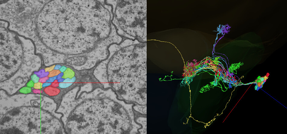
Using the Connectivity app I was able to find at least one T2/T2a, which was connected with an (unfinished) L5 cell: FlyWire. The hidden cells are all the other upstream partners.
I will have to check it for more cells, but thought, it might be useful for other Flyers.
Using the Connectivity app seems to be also a good way to find unfinished cells.
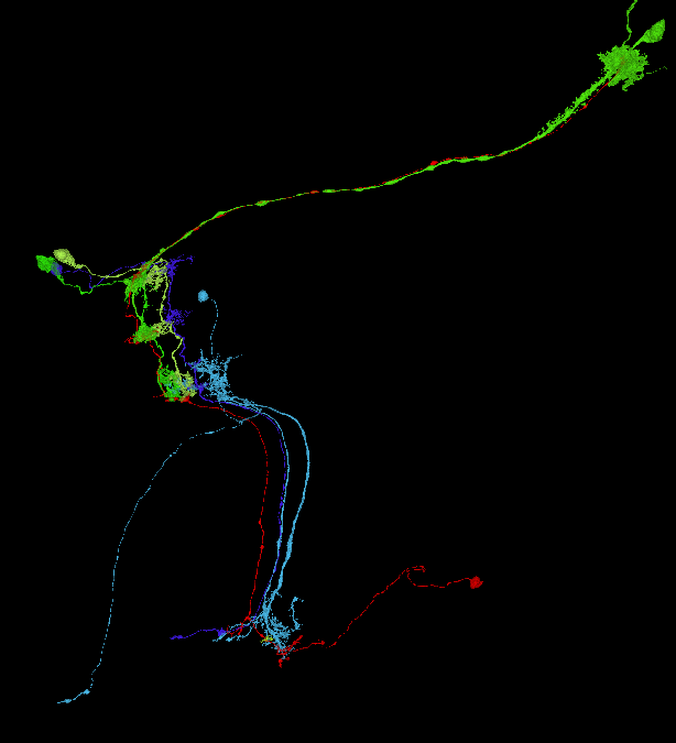
I ran it on our mystery cell and came away with a few insights:
The upper (C-style) portion synapses with an L1;
The medulla bend synapses with Mi1 and a Tm cell (possibly Tm25);
The lobula portion interfaces with a cell type that goes through lobula, medulla, and into the CB.
You can see pathway mirroring very clearly with these cells:
Agree, checking upstream and downstream partners might help. Actually, I have 15 of that kind of cells, that goes through medulla, lobula and projects into the CB. Don’t have a type for them, but checking their synaptic partners might help narrowing down, what that mysterious cell is or isn’t.
For something completely different: does anyone have a foolproof way to differentiate subtypes of T4 and T5? I was trying to find some barriers between LOP layers in 2D, but to no effect. I was also searching for upstream and downstream partners, but it seems, that all the subtypes connect with the same types of cells.
By the way, T4a, T4b, T5a and T5b can be further divided into T4ai and T4aii, T4bi and T5bii, etc. However, the distinction is based only on neurons response (directionality), so it’s impossible to say, which one is which from the anatomy only.