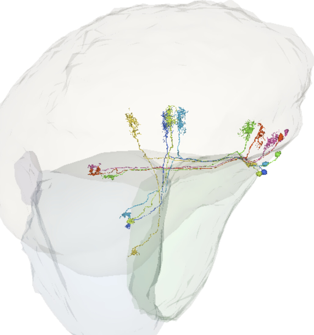
I was thinking about it. My T3 have its proximal end in the lobula neuropil, but I’m starting to think, that the 3D models of the neuropils aren’t exactly matching the 2D slides.
Yesterday I was sure, that they are correct, but the more time passes, the more I think, I got it wrong and the more I agree, that T3 should look only like the ones you posted.
At least, I didn’t post my classifications yet.
Agree about the neuropils not completly matching, making it even more difficult with the T4 and T5, from what i have observed i think there are many T4 cells that are just under the medulla neurophil, because of this i tend to classify all of the ones i am unsure about as T4
I wish, I could make the models more accurate and even add all the layers. Unfortunately, I don’t know how to.
Thanks, for straightening my knowledge about the T3s
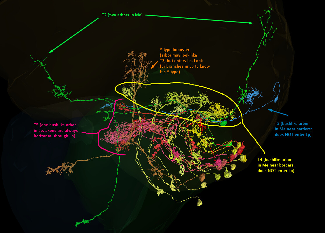
T2 (Green): always has TWO arbors in the medulla. Sometimes the first arbor will look similar to a T3 arbor, sometimes (like these examples) they will be more loose.
T3 (Blue): always has arbor in the medulla near the neuropil borders. Usually it is bush like but it can look a bit taller (tree-like). It NEVER enters the Lobula Plate.
Y Imposter (orange)!: Has an arbor in the medulla near neuropil borders. It goes through Lp and Lo. Branches in the Lp tell you it’s a Y type, not T type.
T4 (Yellow): Has arbor in medulla near neuropil borders, always small bush-like. Does NOT enter Lo.
T5 (Red): Has bush-like arbor in Lobula near borders. Axons always go horizontal through lobula plate (Lp).
Yes, despite the weirdness of the 3D model, those are T4s!
I haven’t been able to find a way to easily tell via 2D where Lp ends and Me begins, without utilizing other known neurons to judge by. But because these are “U-turn” types, and the arbor is close to the neuropil border, it’s safe to assume it’s a T4 type (Lpi do not have a U-turn structure).
I have trouble identifying T2 vs T2a cells. I know the difference in the number of arbors on the Medulla’s branch, but it isn’t always a clear difference. So I found this:
T2a lacks inputs from L cells unlike T2, despite its proximal medulla innervation
Using the Connectivity app I was able to find at least one T2/T2a, which was connected with an (unfinished) L5 cell: FlyWire. The hidden cells are all the other upstream partners.
I will have to check it for more cells, but thought, it might be useful for other Flyers.
Using the Connectivity app seems to be also a good way to find unfinished cells.
I ran it on our mystery cell and came away with a few insights:
The upper (C-style) portion synapses with an L1;
The medulla bend synapses with Mi1 and a Tm cell (possibly Tm25);
The lobula portion interfaces with a cell type that goes through lobula, medulla, and into the CB.
You can see pathway mirroring very clearly with these cells:
Agree, checking upstream and downstream partners might help. Actually, I have 15 of that kind of cells, that goes through medulla, lobula and projects into the CB. Don’t have a type for them, but checking their synaptic partners might help narrowing down, what that mysterious cell is or isn’t.
For something completely different: does anyone have a foolproof way to differentiate subtypes of T4 and T5? I was trying to find some barriers between LOP layers in 2D, but to no effect. I was also searching for upstream and downstream partners, but it seems, that all the subtypes connect with the same types of cells.
By the way, T4a, T4b, T5a and T5b can be further divided into T4ai and T4aii, T4bi and T5bii, etc. However, the distinction is based only on neurons response (directionality), so it’s impossible to say, which one is which from the anatomy only.
Yes, each of them ends in one of the four LOP layers. It’s possible to tell the “a” and “d” types, if they are at or near the border of the LOP, but the others is just a guess. I’m still trying to find a way to make it more accurate. I have almost 500 T4 and T5 to be categorized, but still holding from it, because the possible inaccuracy.
I was thinking about adding “walls” of annotations, if the dividers between the layers were visible in 2D. Unfortunately, no matter the zoom level and the angle of slicing, I couldn’t find any internal boundaries.
No, I didn’t notice such differences.
Here are all my T4 and T5 cells: FlyWire (warning: 492 cells). You can kinda see the layers in LOP, but some of the axonal terminals are between those layers, and it make things harder to guess.
I’ll try to search again in 2D, because I think, the layers have more terminals, why in the boundaries there are more branches.
that was quite a challenge, i guess try to pick out layer for layer, even if you get more than 4 layers maybe easier to see when you get less cells at once, but do not have a good solution
Either there are several types in that bunch or someone have done a bad job when tracing them since they are so different. also several completed cells missing soma.
I guess if you write down a list of id and cordinates on the ones that are identical it could be mass classified by a scientist later.
There are also about 10% of unfinished cells.
I believe, they are of the same type, probably some complex columnar visual projection neurons, but I didn’t find any more info about such cells and I dig through a lot of papers today.
And I believe, they are of the same type, because they all end in the same glomerulus. It’s a typical thing for all the visual projection neurons (e.g. LC, LPLC, LLPC, etc.). The cells themselves can look quite different, but if they go through the same neuropils and end in the same glomerulus, they are usually of the same type.
There are no internal boundaries of the LOP layers - the four layers that we have are based on the location/strata of terminals of neurons. Basically LOP1 is defined by T4a/T5a terminals, etc.
I have been quickly classifying things as putative T4/T5 type because of that: it is easier to classify the types more specifically when you have a comprehensive look at the anatomy, and then even further based on upstream/downstream connections, etc.
There’s a lot of reading to be had about these, but as I was researching this here I found a useful diagram that indicates that the ability to identify these by their dendrite orientations:
(B) Individual T4 dendrites labeled with tdTomtato and Da7::GFP; subtypes a and d pointing in their natural orientation in visual space coordinates (A = anterior, p=posterior, D = dorsal, V = ventral). White arrows indicate preferred directions for every subtype and the dendrites’ proximal (Prox.), central (Cent.) and distal (Dist.) areas are labeled (scale bar: 2 mm). Yellow circle labels first branching point of the dendrite. (C) Individual T5 dendrites labeled with tdTomtato and Da7::GFP; subtypes a and d pointing in their natural orientation in visual space coordinates (A = anterior, p=posterior, D = dorsal, V = ventral). White arrows indicate preferred directions for every subtype and the dendrites’ proximal (Prox.), central (Cent.) and distal (Dist.) areas are labeled (scale bar: 2 mm). Yellow circle labels first branching point of the dendrite.
So it seems that the first line of identifying subtypes here would be by their axon terminals, where obvious; when not obvious, refer to the dendrite cluster’s orientation.