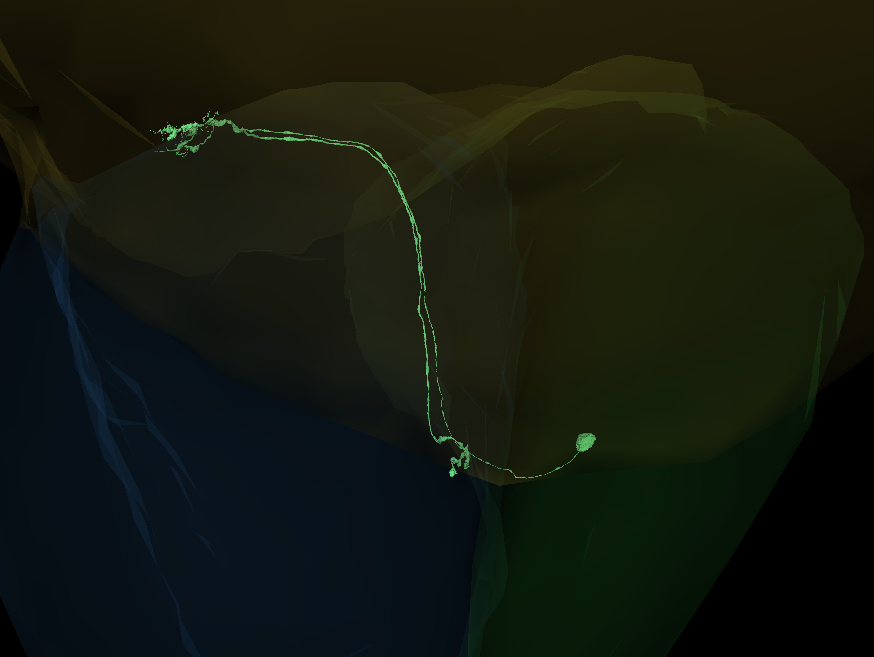
Interesting. With the CBs in these places, they don’t look anything like Dms. They don’t look like anything from Fischbach.
1 Like
i’ve completed cells like these before, if it wasnt for the fact that they are missing the ‘umbrella’ like boutons at the end of the axonal stems theyd look a lot like LAWFs, they may be and be missing the final boutons, didn’t check at the end of the axons. But, in the past ive completed 2-3 of what i thought originally would be LAWFs but they ended up having a singular bouton at the end of the axon vs the umbrella like or grape like structure. Idk maybe theyre one of the cells we dont have examples/pictures of in the gsheet tab? Or a new cell type/subtype not recorded in Fischbach?
EDIT: Checked both axons end in singular boutons, the one on the left is missing a segment but then end of bouton is outside of the tracable dataset.
1 Like
yes, Lawf are the closest ones with the CB being somewhere in the middle of the cell. But they usually have much more synaptic terminals at both ends. Maybe that’s how they look like, when they are near the edge of the medulla.
1 Like
Yeah theyre missing said synaptic terminals (the umbrella lol) but otherwise theyre quite similar, maybe a new subtype.
1 Like
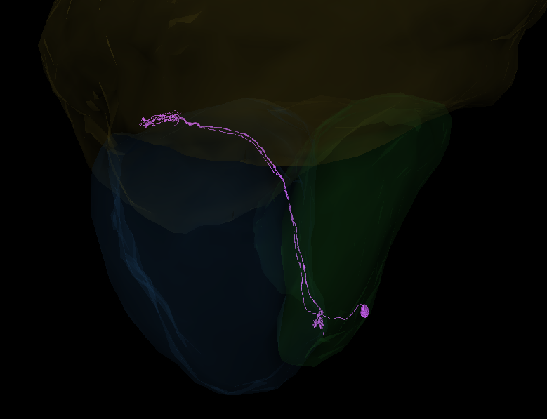
Thank you for finding the cb and removing the merger (i was really unsure if it belonged or not)
agree that it is probably some type of lawf
1 Like
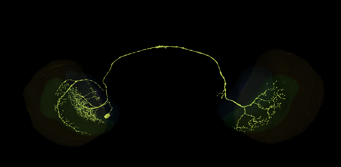
Any idea what these dual hemisphere neurons are? Finding I interface a lot with them in proofing through the lobula plate.
https://ngl.flywire.ai/?json_url=https://globalv1.flywire-daf.com/nglstate/5863790918762496
2 Likes
Those are called bilateral neurons, in general. But there are 394 entries for “bilateral” on FlyBase.org in “Anatomy Ontology” section for the Drosophila alone.
Part of the neuron looks a little bit like an Lpt2, but those are unilateral, so there’s that.
1 Like
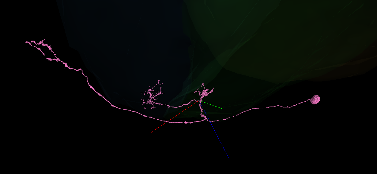
Here’s another one I don’t recognize from the Lobula Plate area. It has dendritic branches in both the lobula plate and lobula, with boutons heading into the central brain.
Edit: Updating link with a second similar structure
https://ngl.flywire.ai/?json_url=https://globalv1.flywire-daf.com/nglstate/6198215644807168
3 Likes
Might be one of the LLPC or LPLC types.
3 Likes
Yep you’re right, after digging about it seems it’s LLPC, most likely LLPC1. Thank you!
4 Likes
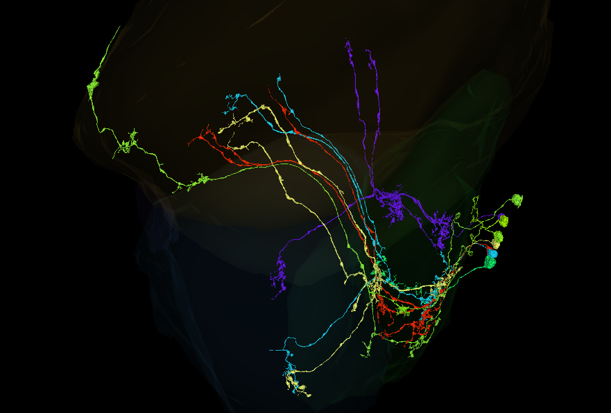
I have 8 cells here - FlyWire - from the outer lobula plate rind that I was not able to complete or identify in my pass (as described here). I would love your assistance!
I think a few of these are probably a Y type, but there’s confusing things all around here. One is probably centrifugal.
Feel free to make live edits if you find something!
2 Likes
I’d say, 4 of them are Y.
The short green one (065) might be an LPi.
The longest could be a merger (didn’t found any merging spot, though). The outer part has a missing extension, that looks like a C cell (I didn’t trace it to the end).
The one ending with 326 is unfinished. It’s continuation goes into a merger, but I didn’t check, what part of that merger is correct and if it continues beyond that, so it’s hard to say, for now, what type it is.
I don’t know, what to think about 218, but it also looks, like its unfinished.
2 Likes
Thanks for the feedback! I’ve done some updates and narrowed things down a bit:
- Marked the four y cells as Putative Y and sorted them out. I had forgotten about Y11 and Y12 types which match them a lot more.
- Edited two cells, one of which I’ve recompleted as Lpi type (720575940622000372, removed from link below). The other (-629 in the link) has been fleshed out a little more but I still feel unsure about it being complete, although it is probably Lpi too.
- (-326) definitely feels like a merger, and I’ve added the opposite trace-back soma. I think it’s somewhere along the ‘break’ in the 3D, but I can’t put my finger on it still.
- (-740), the extra long one, I haven’t been able to find anything more to point to a merger or an extension. The medulla end definitely reminds me of C-types but the rest of the structure doesn’t add up to anything. My best guess is that this is an LLPC that is merged with a branch that heads up to the medulla, and is missing its branch to the central brain.
Updated link: FlyWire
1 Like
https://ngl.flywire.ai/?json_url=https://globalv1.flywire-daf.com/nglstate/5203824817995776
- Added the correct extension to -326. It’ll be a T cell after merging,
- I think, the -629 looks complete enough,
- Added more segments to -740. Now the top part definitely is a C cell. For now, I couldn’t find a merger either. I’m going to go to sleep now, so I’ll look at it tomorrow.
2 Likes
I’ve checked (more or less) the whole -740 and didn’t find any mergers. I’m starting to think, maybe there isn’t a merger. Maybe it is a C cell, but malformed. Maybe it grew in the wrong place at the wrong time and was dragged away from its normal position. Not sure, if it’s possible though ![]()
2 Likes
Thanks so much for the assistance and feedback, KK!
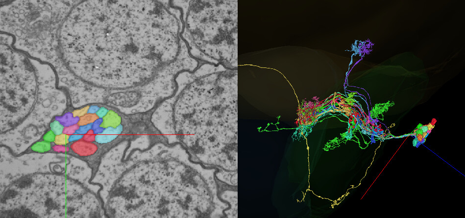
I’ve eliminated the T cell and Lpi now, which leaves us just with this stranger. I’ve merged together your additions, and made a few clean ups/additions myself where I could find them. I also combed through the 2D again, and I’ve marked two points that might be mergers. Though I will say even if they are mergers, we’re still drawing a blank on what this cell is!
https://ngl.flywire.ai/?json_url=https://globalv1.flywire-daf.com/nglstate/4988991979913216
I’ve tried looking at friend cells in different places, but not finding any clues. This bundle is mostly T5s, a few T4s, and one Tlp. I’m most inclined to think it’s an Lpi merged with something else, assuming it isn’t, as you suggested, a poorly grown ‘mistake’ neuron.
2 Likes
You’re welcome ![]()
Unfortunately, I can’t find any mergers either.
Maybe it’s a new kind od cell, some sort of probe connecting lamina with lobula and lobula plate. There aren’t any other cells directly connecting these neuropils, so maybe there’s a C cell repurposed just for this.Who knows. After all, we’re doing researching, so might find unknown unknowns ![]()
2 Likes
Quick guide, how to differentiate between T cells:
T1: Lamina → Retina
T2: Medulla → Lobula (goes through the whole Medulla)
T3: Medulla → Lobula (arborizes in the proximal part of the Medulla only)
T4: Medula → Lobula Plate
T5: Lobula → Lobula Plate
ChaTnew1: Medulla → Lobula and Lobula Plate
The last one is also known as Tnew1 in some pictures.
Categorizing T cells definitely needs to have the optic lobe’s neuropils turned on, because they sometimes have their arborizations very close to the borders of the neuropils.
1 Like
do you have a picture of the ChaTnew1 have not seen this before?
i am thinking on the differences more like this
T1= basket
T2= long arbour
T3= short arbour
T4 = turn 180 degree, goes into medulla
T5= turn 180 degree, goes into lobula
I was sure, that Tnew1 (ChaTnew1) cell has already been posted in the Visual Cell Type Illustrations thread. Turns out, it wasn’t, so I’ve added it.
Edit: The part below is wrong
as for T3 and T4 it’s not so easy, unfortunately.
For example:
T3:
https://ngl.flywire.ai/?json_url=https://globalv1.flywire-daf.com/nglstate/5662774554263552
T4:
https://ngl.flywire.ai/?json_url=https://globalv1.flywire-daf.com/nglstate/6733026448900096
There’s also a possibility, to mistake T4 with T5.