I’m starting to think, that those “Mt” cells from one of the previous posts are actually Pm2s, and the cells, we identified as Pm2 in the sheet are some type of an Mt (it’s hard to find any images of the Mt cells).
There are a couple of things, that make me think that:
All the other Pms are only inside the medulla.
Only the Pm4s have a bunch of parallel neurites - both Pm1 (and Pm1a) and Pm3 have single neurites.
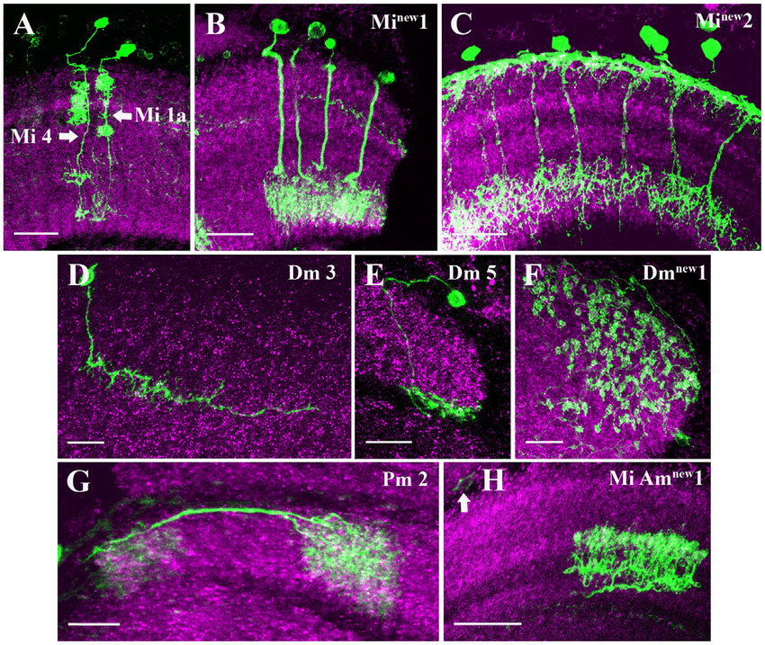
The image in the Fischbach’s paper shows a Pm2 cell, that have clearly visible branches over (distal to) the synaptic terminals, and the terminals themselves are sparser and more blob-like, than the cells from the sheet.
The image here shows a Pm2 cell, that has sparser arborization. The image also shows something, like a second arborization, but it might be a part of a different cell (I’ve seen such cases in a couple of other photos of this type). The description below the picture also says only about the arborization in the medulla and doesn’t mention about anything else.
The description here also talks only about arborization in the medulla.
I’ve found at least one cell of the “new” type, that has been identified as a Pm2 by one of the researchers.
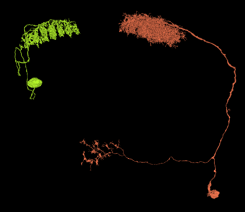
The yellow-green cell on the left - I think, it might be a Pm2 cell
The coral-orange on the right - the type, that we’ve identified as a Pm2, but might be an Mt
1 i can see the difference with the other Pm only beeing in medula, but in the fishback paper the steam cut of before it reaches a cb, to me that might suggest the cb is somewhere outside/far away.
2 i have no idea how you can say that Pm 1 and 3 have no parellell branches, yes not every branch in a bundle like Pm 4 but still they run paralell after the split for a good while before branching of. Yours cells split of at a angle like most of the other celltypes also do except PM and Lai (possible others i cant remember)
3 i find that the pictures from fishback is not very good too identify with always could ofc be some other cells type. but yours are still not a Pm2 imo
4 and 5 maybe difficult too see the second arbor in the earlier articles? or could ofc be some merger in the steam on the example
6 everyone can do mistakes
I think the “new” type is either a Mi 11 in the fishback paper you even have that loop on the steam as that example cell without that beeing any evidence, or some other Mi type. The other choice is a Mt cell
ad. 1. The (assumed by me) Pm2 have their soma in one of the two narrower ends of the medulla rind. I think, that could be a big problem in the method Fischbach used to analyze the cells, hence the missing CBs on their pictures.
ad. 2. In Pm1 and Pm1a there’s almost no paralelism. There’s little more in Pm3, but still not so prominent. Also, when doing microscophic imaging, the paralelism isn’t very visible (I suppose), so that characteristics my not be a reason for a cell to be associated with one or another group.
ad. 6. I’ve seen at least 3 other researchers identifying this kind of cell as a Pm2. I’ve also asked at Slack and the person, that responded to me, thinks this cell might indeed be a Pm2 and the other one - not so sure.
As for Mi 11, I think these or these are better candidates.
I’ve also dig through a ton of papers looking for some info (especially, pictures) of the Mts, but hadn’t have much luck. The only ones I’ve found, are the ones, that are already in the “Visual…” thread on the forum. So any new info on that topic would be very useful.
I guess it is yours call if you want to mark them as Pm 2 or not, too me it is difficult to say that is a Pm 2 but understand that you think it.
What do you plan doing with it beeing two layers?
I really wish we could group the cells in the offisical search feature so it would be possible to mark the cells as belonging too this group, so if needed the whole group could be classified again if needed and lose the “wrong tag” but suppose the spreadsheet works like that.
Agree, renaming groups of cells would be great. Currently we have a situation, when the wrongly assigned labels are still shown in the search results even, when they have already been correctly labeled later.
As for the two layers, I’m currently holding myself from identifying them as anything. I have a file, where I keep IDs of all cells in different groups with some descriptions that characterize them in various ways. I have maybe 5 or 6 such groups. I’ll hold with them until a new info will be found. In the meantime, I’m working with the basic Mi cells (currently Mi1, yesterday I did many Mi4).
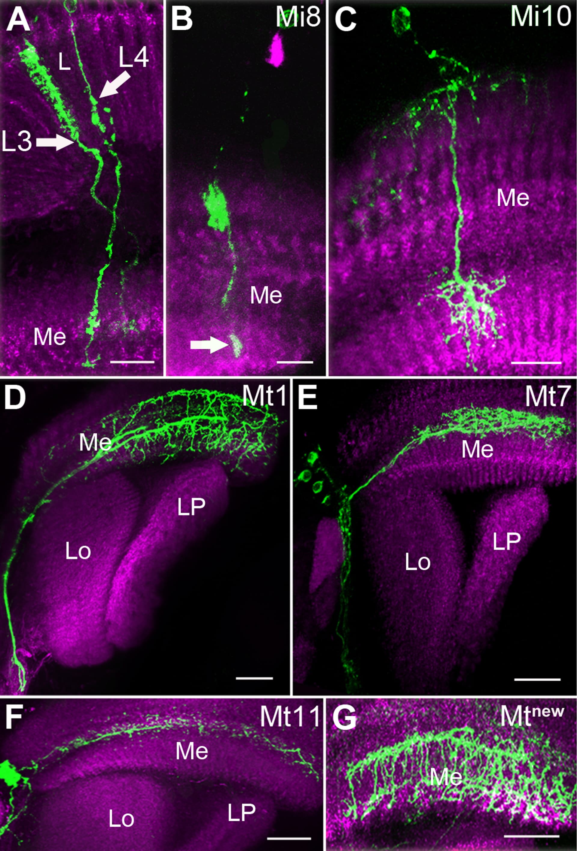
First problem i am going in the direction of Mi8 from Fishback just because i think the shape look closer (even if the drawing in fishback is not the best) it looks like Mi9 is on layer down compared to 8 and i think we would have been able to see this layer differance having so many cells.to compare if both was in there. And the number of cells are also pretty close to what i would expect from a layer of Mi. As far as i know we have not been able to find any examples on Mi9 (same type classified as both Mi8 and Mi9)and I am starting to wonder if they could have made a mistake using cells from different part of medulla and thus seeing it as different layers? Or if there are actually a Mi9 type also.
Thanks! Great explanation, why would there be the two types. Seeing, that both the more distal and the more proximal arborizations are shifted by one level, that could definitely happen with the curvature of the medulla.
Second problem
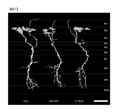
First link looks like what is classified as Mi 13 by Marion Silies with referance too Takemura et al. 2013) I have not been able to find this paper as open source to check it. From what i can see in Fishback the cell classified as Mi12 have a much broader first arborisation and it also looks like it have thinner branches. Also this cell FlyWire was in your layer of Mi 8 but do probably belong in this layer of Mi 13?
As for the second link at first look i am also thinking Mi10, but looking closer i am thinking it might be a unknown Mi cell or alternativly a bad drawing in fishback. The differances i see is that the first arbor looks mainly horisontal in fishback while this cell goes more vertical also. The last arbour is supposed to be in layer 9 while this cell goes to layer 10 FlyWire of course all of this could be based on having lower quality images in the first classification, but the sum of differences is a bit too much for me.
As for the Mi(new)2 i agree that this looks very close too Mi 12 as drawed in fishback
Maybe not the correct thread for this, but i’ve started IDing L1’s (they tile nicely in line with the Dm4’s ), and there seem to be a few that could be mistaken for what i would have been calling glial before (the ones with colour here, for example:)
https://ngl.flywire.ai/?json_url=https://globalv1.flywire-daf.com/nglstate/6254882470232064
Obviously they arent glial cells, but they are a bit more difficult to separate from surrounding similar structures and/or real glials. Is there a better (or correct) term i could use to refer to them? (and maybe a question for GM’s, if not answered somewhere already: should i put any extra info in the ID to differentiate between these and the ‘normal’ ones?)
Very interesting, Imo they probably are L1, but i suppose you could mark them out in some way like copy them out too a separate tab too see if there is any systematic differanse or if it might just be some colouring error in that one area?
Hi there! I would just label them as L1. As annkri said, it could just be an error in the dye/coloring/membrane burst, etc. that is causing the more glial-like spread. As whole, the structures are L1 cell type like the others.
Here is (I hope!) an open source link to the Takemura et. al 2013 paper: Europe PMC. I think folks will find the Supplementary Material #2 of great interest. It’s a Word Doc you’ll have to download, but it has some good images.
some great pictures you really should add some of them to the optic lobe cell name guide, so we have everything in the same place. i am thinking page 1-6 and 12-18 and 35-47 is the ones that would be most helpfull. I feel that the rest we either have good control on based on what we already have or too little of the cell is in medulla.
Thanks @Amy_R_Sterling!
In the mean time, I’ll be working on the other cells from similar groups (still have to identify them).
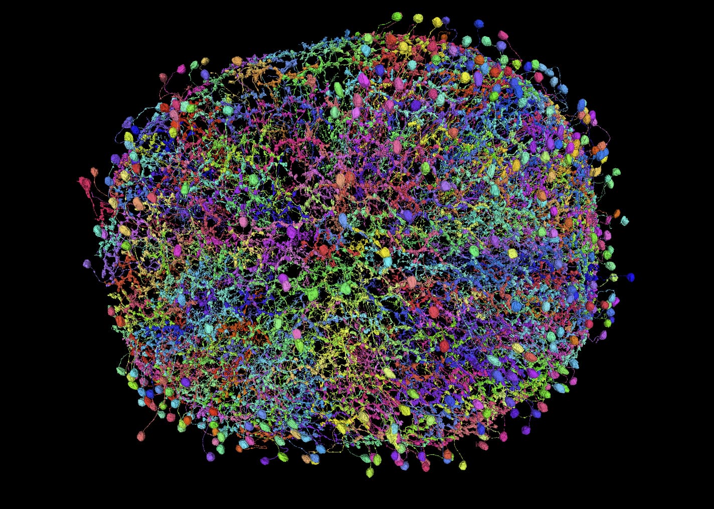
And yes, both these and Lawf cells create really nice nets, showing where all the cartridges are. It also helps with finding the missing ones, where the net doesn’t seem to be complete.